. 2022 Aug 30;13(1):5091.
doi: 10.1038/s41467-022-32054-0.

Organic phosphorescent nanoscintillator for low-dose X-ray-induced photodynamic therapy

Affiliations

Organic phosphorescent nanoscintillator for low-dose X-ray-induced photodynamic therapy

Xiao Wang et al. Nat Commun. .

Abstract

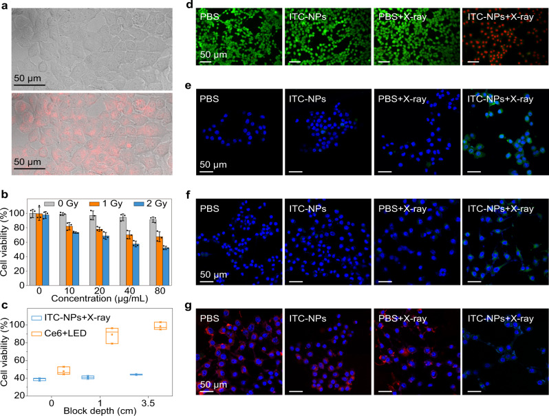
X-ray-induced photodynamic therapy utilizes penetrating X-rays to activate reactive oxygen species in deep tissues for cancer treatment, which combines the advantages of photodynamic therapy and radiotherapy. Conventional therapy usually requires heavy-metal-containing inorganic scintillators and organic photosensitizers to generate singlet oxygen. Here, we report a more convenient strategy for X-ray-induced photodynamic therapy based on a class of organic phosphorescence nanoscintillators, that act in a dual capacity as scintillators and photosensitizers. The resulting low dose of 0.4 Gy and negligible adverse effects demonstrate the great potential for the treatment of deep tumours. These findings provide an optional route that leverages the optical properties of purely organic scintillators for deep-tissue photodynamic therapy. Furthermore, these organic nanoscintillators offer an opportunity to expand applications in the fields of biomaterials and nanobiotechnology.


Conflict of interest statement

The authors declare no competing interests.

Figures

Fig. 1
Fig. 1. Schematic representation of X-PDT based on organic phosphorescent nanoscintillators.
a Process to prepare organic nanoparticles in aqueous solution and subsequent 1O2 generation under X-ray irradiation. Specifically, an organic scintillator 9,9’-(6-iodophenoxy-1,3,5-triazine-2,4-diyl)bis(9H-carbazole) (ITC) and PEG-b-PPG-b-PEG (F127) were dissolved in chloroform. The resultant solution was evaporated and dissolved in aqueous solution accompanied by intense sonication to obtain organic nanoparticles. Under X-ray irradiation, electrons (orange circles) are mainly ejected from the inner shell of heavy atoms in ITC (step 1), generating massive electron-hole pairs (step 2). The following charge recombination produces singlet and triplet excitons, in a certain ratio. With the assistance of enhanced intersystem crossing (ISC) owing to strong spin-orbit coupling (step 4), a large amount of triplet excitons is produced. The resultant triplet excitons excite 3O2 to yield 1O2 by triplet-triplet annihilation (TTA, step 5). b Illustration of the mechanism in the treatment of deep-seated tumours in vivo. The local 3O2 molecules absorb energy to produce massive 1O2. The resultant 1O2 species lead to membrane oxidation and mitochondrial damage, which together effectively kill cancer cells.
Fig. 2
Fig. 2. Characterization and photophysical properties of organic phosphorescent nanoscintillators in aqueous solution.
a TEM image of the prepared nanoparticles (scale bar: 500 nm). b Size distribution of nanoparticles obtained by TEM. c Steady-state photoluminescence (PL, dashed line) and time-gated phosphorescence (Phos., solid line) spectra of the nanoparticles in solution, respectively (top). And the radioluminescence (RL) spectrum of the nanoparticles in solid state (bottom). d Time-resolved decay profile of emission wavelength at 530 nm of the nanoparticles under excitation at 340 nm. e Absorption spectra of the aqueous ITC suspension in the presence of anthracene-9,10-diyl-bismethylmalonate (ADMA) under different X-ray irradiation doses (X-ray dose rate: 2.8 mGy/s, time interval: 72 seconds). f Dose dependence of the absorbance variation (monitored at 379 nm) of the mixture of ITC (or anthracene) suspension and ADMA in water.
Fig. 3
Fig. 3. Evaluation of organic phosphorescent nanoscintillators for X-PDT in vitro.
a Confocal microscopic images of 4T1 cells after incubation with ITC nanoparticles (ITC-NPs). The red colour (bottom) is from the phosphorescence of the ITC-NPs, indicating effective cell uptake, as it appears in the same position as the bright-field images of the 4T1 cells (top). b Viability of 4T1 cells incubated with different concentrations of ITC-NPs. Note that each group was treated parallelly with or without X-ray irradiation (1 or 2 Gy). The statistical data are expressed as mean values ± S.D. (n = 3 independent experiments). Note that the total dose that medium and cells irradiated is 1-2 Gy. c Chicken breast block depth dependence of viability of 4T1 cells incubated with ITC-NPs (X-ray: 2 Gy) or Ce6 (LED light: 670 nm, 300 mW/cm2, 90 J/cm2, 300 s). Median and interquartile ranges are presented for the box plot (n = 3 independent experiments). d Calcein-AM/PI co-staining assay of 4T1 cells after treatment with PBS, PBS + X-rays, ITC-NPs, and ITC-NPs+X-rays (X-ray irradiation: 2 Gy) (Green: live cells; Red: dead cells). e Confocal laser scanning microscope (CLSM) images of 4T1 cells stained with SOSG after incubation with PBS or ITC-NPs. Two treatment groups were subjected to X-ray irradiation (2 Gy). f CLSM images of lipoperoxides in 4T1 cells after incubation with PBS or ITC-NPs with or without X-ray irradiation (2 Gy). Green fluorescence indicates lipid ROS formation after staining with BODIPY-C11. g CLSM images of the changes in the mitochondrial membrane potential of 4T1 cells irradiated with or without X-rays (2 Gy). Red fluorescence indicates positive membrane potential, whereas green fluorescence indicates decreased membrane potential.
Fig. 4
Fig. 4. Assessment of organic phosphorescent nanoscintillator for X-PDT in vivo after intravenous injection.
a Tumour growth curves of 4T1 tumours with different treatments (****P < 0.0001, ***P = 0.0003). On day 0, 0.4 Gy X-ray irradiation was given without further treatment. The statistical data are expressed as mean values ± S.D. (n = 5 biologically independent animals). Statistical significance was assessed via unpaired two-sided Student t-test. b Tumour weights after different treatments for 14 days. The inset shows the resulting tumour photographs. Median and interquartile ranges are presented for the box plot (n = 5 biologically independent samples). c H&E-stained images of tumour slices after various treatments.

References

    1. Zhang C, et al. Marriage of Scintillator and semiconductor for synchronous radiotherapy and deep photodynamic therapy with diminished oxygen dependence. Angew. Chem. Int. Ed. 2015;54:1770–1774. doi: 10.1002/anie.201408472. - DOI - PubMed
    1. Kamkaew A, Chen F, Zhan Y, Majewski RL, Cai W. Scintillating nanoparticles as energy mediators for enhanced photodynamic therapy. ACS Nano. 2016;10:3918–3935. doi: 10.1021/acsnano.6b01401. - DOI - PMC - PubMed
    1. Lu K, et al. Low-dose X-ray radiotherapy–radiodynamic therapy via nanoscale metal–organic frameworks enhances checkpoint blockade immunotherapy. Nat. Biomed. Eng. 2018;2:600–610. doi: 10.1038/s41551-018-0203-4. - DOI - PubMed
    1. Ni K, et al. Nanoscale metal-organic frameworks for mitochondria-targeted radiotherapy-radiodynamic therapy. Nat. Commun. 2018;9:4321. doi: 10.1038/s41467-018-06655-7. - DOI - PMC - PubMed
    1. Sun W, et al. Monodisperse and uniform mesoporous silicate nanosensitizers achieve low-dose X-ray-induced deep-penetrating photodynamic therapy. Adv. Mater. 2019;31:1808024. doi: 10.1002/adma.201808024. - DOI - PubMed

Publication types