Review
. 2023 Feb;21(2):97-111.
doi: 10.1038/s41579-022-00780-3. Epub 2022 Aug 30.

Staphylococcus epidermidis and its dual lifestyle in skin health and infection

Affiliations
Review

Staphylococcus epidermidis and its dual lifestyle in skin health and infection

Morgan M Severn et al. Nat Rev Microbiol. 2023 Feb.

Abstract

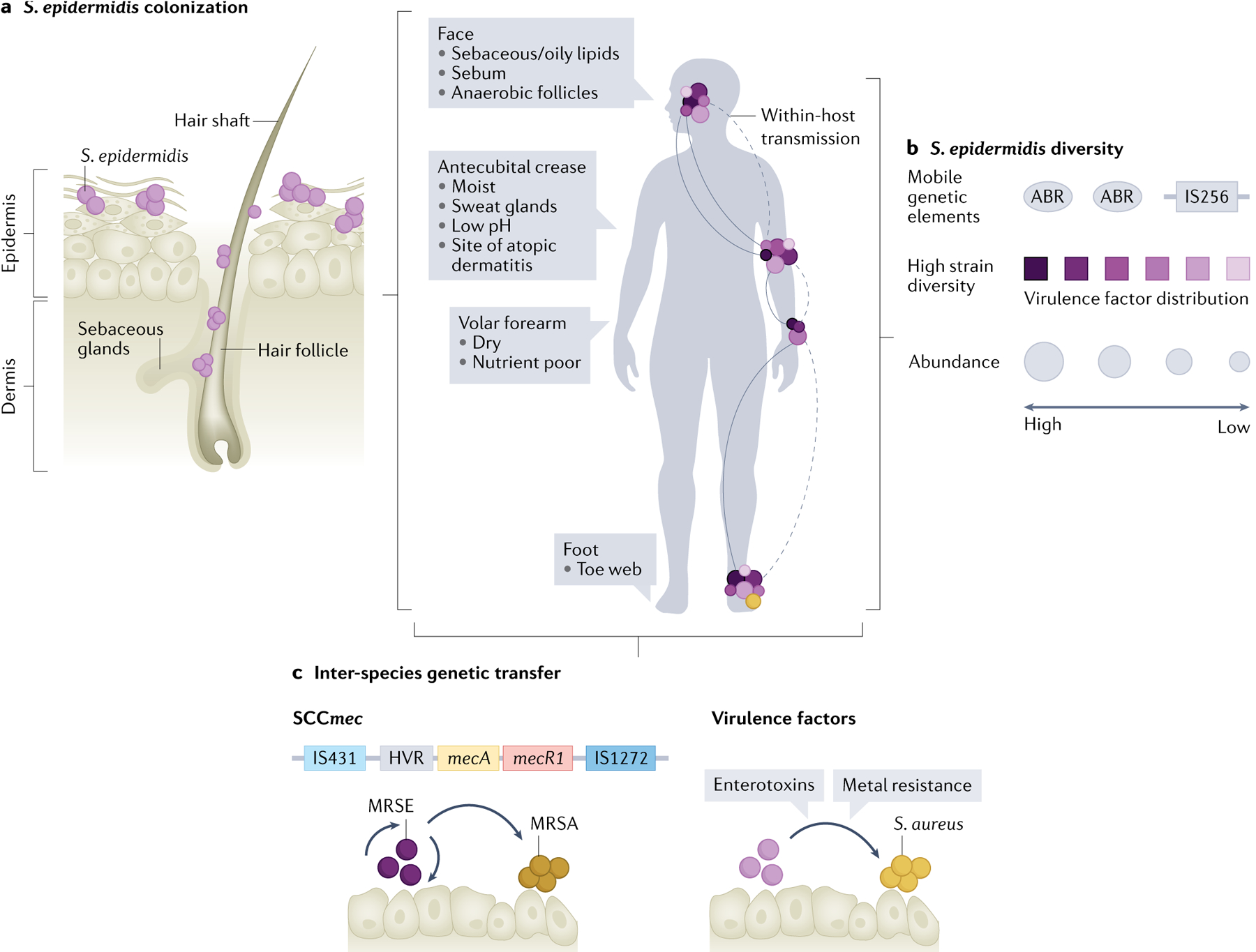
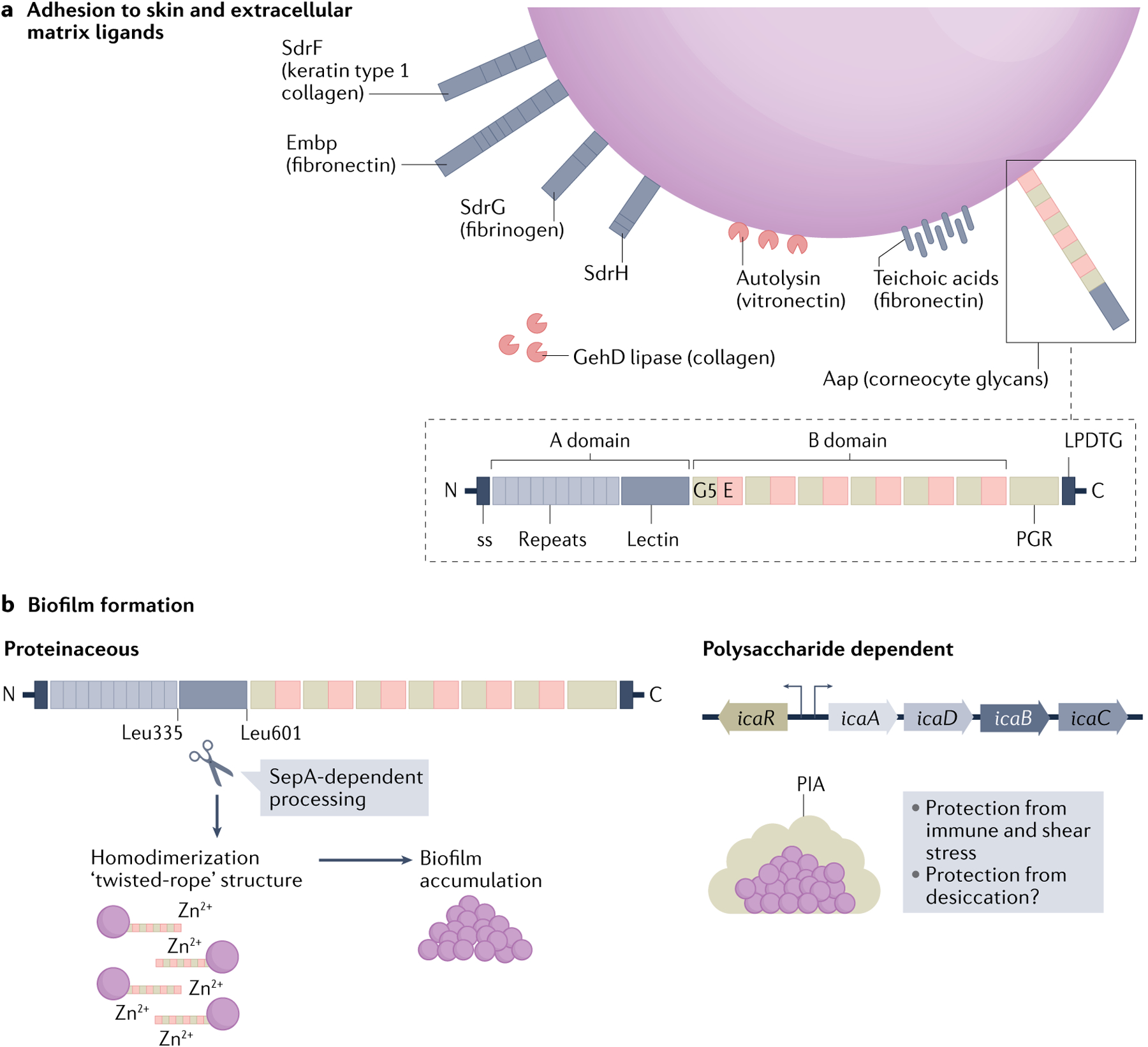
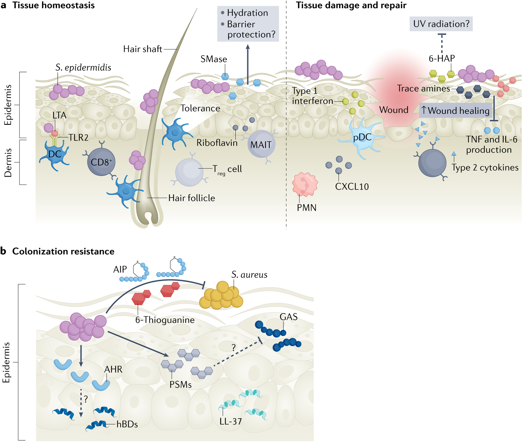
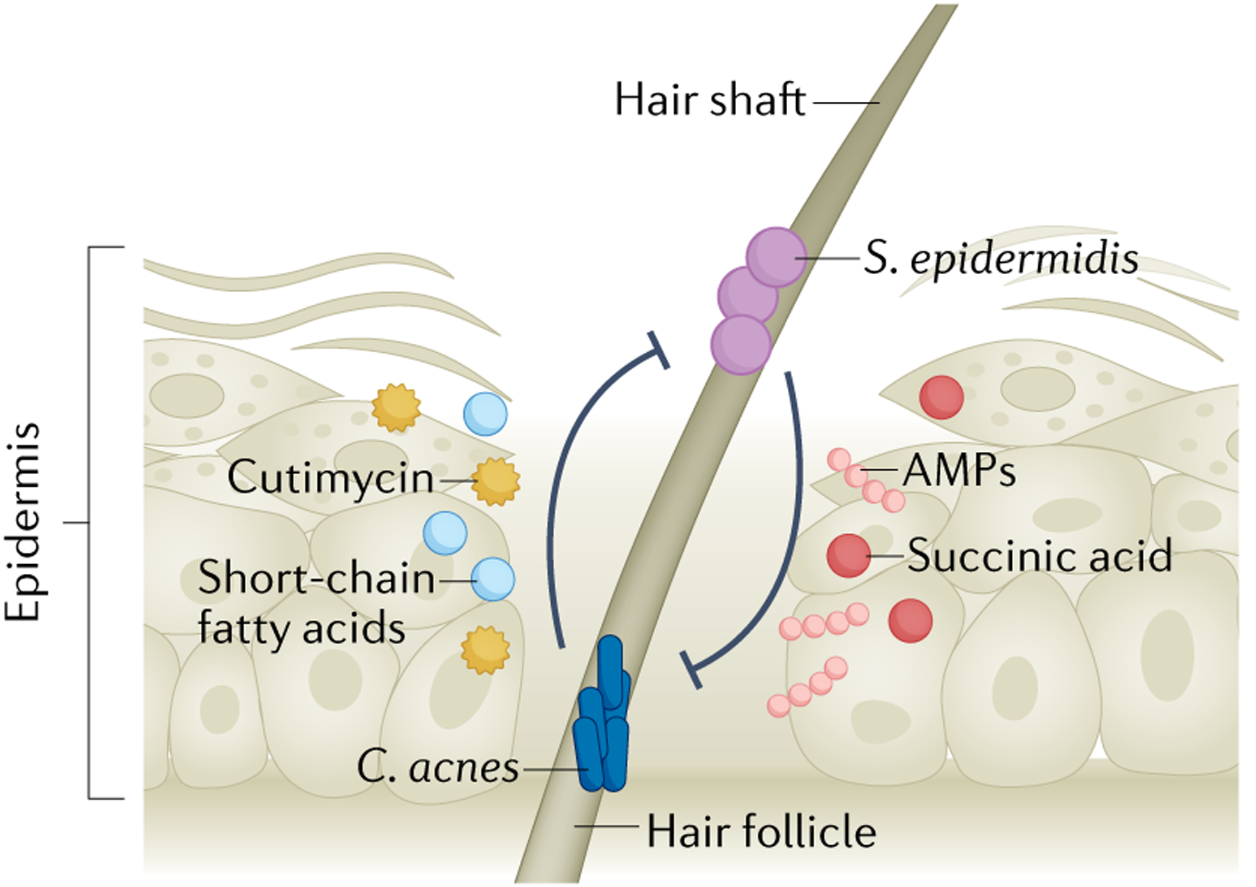
The coagulase-negative bacterium Staphylococcus epidermidis is a member of the human skin microbiota. S. epidermidis is not merely a passive resident on skin but actively primes the cutaneous immune response, maintains skin homeostasis and prevents opportunistic pathogens from causing disease via colonization resistance. However, it is now appreciated that S. epidermidis and its interactions with the host exist on a spectrum of potential pathogenicity derived from its high strain-level heterogeneity. S. epidermidis is the most common cause of implant-associated infections and is a canonical opportunistic biofilm former. Additional emerging evidence suggests that some strains of S. epidermidis may contribute to the pathogenesis of common skin diseases. Here, we highlight new developments in our understanding of S. epidermidis strain diversity, skin colonization dynamics and its multifaceted interactions with the host and other members of the skin microbiota.


Conflict of interest statement

Competing interests

The authors declare no competing interests.

Figures

Fig. 1 |
Fig. 1 |. The commensal lifestyle of Staphylococcus epidermidis.
a | The skin can be subdivided into different ecological niches based on physio-chemical properties such as density of sebaceous and sweat glands, level of moisture and oxygen tension. Staphylococcus epidermidis colonizes all these environments at different densities, including the outermost stratum corneum as well as the interior of the hair follicle, and is a dominant colonizer of moist sites. b | Recent metagenomics studies revealed high strain-level heterogeneity of skin-colonizing S. epidermidis (represented by various colours) (BOX 1). These strains share genetic content, including virulence factors and antibiotic resistance factors (ABRs), via horizontal gene transfer of mobile genetic elements, including plasmids, phages and transposable elements (such as the insertion sequence IS256). Body sites are colonized by varying levels of S. epidermidis (indicated by circle size) with the highest density of S. epidermidis in moist or sebaceous sites. c | Horizontal gene transfer of the staphylococcal cassette chromosome mec (SCCmec), which encodes resistance to methicillin, between S. epidermidis and Staphylococcus aureus. Other virulence factors can also be exchanged between these organisms, including enterotoxins and genes for metal resistance. MRSA, methicillin-resistant S. aureus; MRSE, methicillin-resistant S. epidermidis.
Fig. 2 |
Fig. 2 |. Staphylococcus epidermidis adhesion and biofilm formation.
a | Staphylococcus epidermidis binds components of skin (ligands indicated, if known) and the extracellular matrix through expression of surface-anchored proteins, called adhesins. This family of microbial surface components recognizing adhesive matrix molecules (MSCRAMMs) includes several serine aspartate repeat containing (Sdr) proteins. Other non-MSCRAMM cell wall-associated proteins with roles in cellular adhesion include the giant Embp protein, the bifunctional autolysin and the bifunctional lipase GehD. Many S. epidermidis isolates encode the large (140 kDa), multi-domain accumulation associated protein (Aap) that is anchored to the cell wall by sortase via the LPDTG motif in the carboxy terminus. Aap export from the cell is facilitated by the amino-terminal secretion sequence (ss). The A domain is composed of short, 16 amino acid repeats followed by a lectin-like domain. The B domain contains a variable number (5–17) of 128 amino acid repeats of G5 domains (named for their conserved glycine residues) and E spacer regions. The C-terminal proline–glycine rich region (PGR) and the cell wall anchoring motif (LPDTG) are also indicted. Aap specifically mediates attachment to the host stratum corneum through the A-domain lectin, which leads to epithelial colonization. b | Aap also mediates proteinaceous biofilm attachment and accumulation. The metalloproteinase SepA cleaves the N-terminal A domain of Aap at specific amino acid residues (Leu335 and Leu601). Cleavage is followed by homodimerization of the B domain in a zinc-dependent manner. Many, but not all, S. epidermidis strains have the icaADBC operon, which encodes polysaccharide intracellular adhesin (PIA; also known as poly-N-acetylglucosamine (PNAG)), as well as the icaR gene, which encodes a regulatory protein. PIA is composed of repeating β-1,6-linked N-acetylglucosamine residues and its production promotes biofilm formation. Such polysaccharide-dependent biofilms may protect mature biofilms from immune, antibiotic or shear stress. In addition, the polysaccharide has been speculated to aid in protection against desiccation but further investigation is warranted.
Fig. 3 |
Fig. 3 |. Staphylococcus epidermidis mediates skin homeostasis, barrier repair after injury and colonization resistance.
a | Homeostasis: early-life Staphylococcus epidermidis skin colonization in the hair follicle primes the cutaneous immune response to tolerate future commensal colonization via activation of regulatory T cells (Treg cells). S. epidermidis-derived riboflavin is sensed by unconventional mucosal-associated invariant T cells (MAIT cells), priming them to become a dominant type 17 effector subset. CD11B+ dendritic cells (DCs) sense S. epidermidis healthy adult skin colonization and stimulate CD8+ T cell migration. S. epidermidis lipoteichoic acid (LTA) is sensed by dendritic cells via Toll-like receptor 2 (TLR2) signalling. S. epidermidis produces a potent sphingomyelinase (SMase) on human skin that increases ceramide content, thus promoting skin hydration and, possibly, barrier integrity. Barrier repair: S. epidermidis and its molecular products including trace amines, lipopeptide 78 and LTA dampen the inflammatory response (TNF and IL-6 production) to promote wound healing. S. epidermidis-recruited polymorphonuclear leukocytes (PMNs) stimulate plasmacytoid dendritic cells (pDCs) via CXCL10 signalling to secret type I interferons in the wound site to aid healing. CD8+ T cells express type 2 cytokines to promote wound repair. 6-N-hydroxyaminopurine (6-HAP) may also protect skin from tumour development, although further studies are necessary to clarify the mechanism and the 6-HAP biosynthetic pathway (dashed line). b | Colonization resistance: S. epidermidis excludes Group A Streptococcus (GAS) and Staphylococcus aureus from skin. Autoinducing peptides (AIPs) block S. aureus quorum sensing and the purine analogue 6-thioguanine inhibits S. aureus purine biosynthesis. S. epidermidis phenol soluble modulins (PSMs) synergize with host antimicrobial peptides (AMPs) including LL-37 to augment killing of S. aureus and GAS, although the mechanism warrants further investigation (dashed line). Unidentified secreted factors from S. epidermidis signal via TLR2 to stimulate keratinocyte production of human β-defensin 2 (hBD2) and hBD3. Unidentified S. epidermidis factors activate the keratinocyte aryl hydrocarbon receptor (AHR) and increase hBD3 expression. Some strains of S. epidermidis produce AMPs that specifically kill certain pathogenic species.
Fig. 4 |
Fig. 4 |. Staphylococcus epidermidis competes with Cutibacterium acnes in the hair follicle.
Cutibacterium acnes is a dominant hair follicle colonizer, and its expansion is correlated with progression of the common skin disease acne vulgaris. C. acnes encounters and competes with follicle-resident Staphylococcus epidermidis through production of the antimicrobial peptide (AMP) cutimycin. C. acnes inhibits S. epidermidis biofilm formation and sensitizes S. epidermidis to antibiotic killing through production of several short-chain fatty acids. C. acnes strains may produce other AMPs but this remains unclear. S. epidermidis counters this competition through production of AMPs and fermentation of follicle-available glycerol to multiple short-chain fatty acids, including acetic acid, butyric acid, lactic acid and succinic acid, to suppress C. acnes overgrowth. S. epidermidis strains may also utilize an ESAT6 secretion system to compete with C. acnes (not indicated), but this remains to be experimentally determined.

References

    1. Byrd AL, Belkaid Y & Segre JA The human skin microbiome. Nat. Rev. Microbiol 16, 143–155 (2018). - PubMed
    1. Gallo RL Human skin is the largest epithelial surface for interaction with microbes. J. Invest. Dermatol 137, 1213–1214 (2017). - PMC - PubMed
    2. This perspective highlights the previously underappreciated large surface area of skin.

    1. Bay L et al. Universal dermal microbiome in human skin. mBio 11, e02945–19 (2020). - PMC - PubMed
    1. Nakatsuji T et al. The microbiome extends to subepidermal compartments of normal skin. Nat. Commun 4, 1431 (2013). - PMC - PubMed
    1. Scharschmidt TC et al. A wave of regulatory T cells into neonatal skin mediates tolerance to commensal microbes. Immunity 43, 1011–1021 (2015). - PMC - PubMed

Publication types