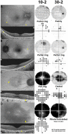
Visual field examinations using different strategies in Asian patients taking hydroxychloroquine
- PMID: 36042337
- PMCID: PMC9427842
- DOI: 10.1038/s41598-022-19048-0
Visual field examinations using different strategies in Asian patients taking hydroxychloroquine
Abstract
In this study, we investigated the patterns of visual field (VF) defects and the diagnostic abilities of VF tests using different strategies in Asian patients with hydroxychloroquine retinopathy. Patients screened for hydroxychloroquine retinopathy using optical coherence tomography, fundus autofluorescence, VF, and/or multifocal electroretinography were included. The VF was performed using the Humphrey 30-2 and/or 10-2 strategy, and 2,107 eyes of 1,078 patients with reliable results, including 136 eyes of 68 patients with hydroxychloroquine retinopathy, were analyzed. The characteristics of VF findings were evaluated and the sensitivity and specificity were compared between the 30-2 and 10-2 tests in subgroups of retinopathy severity and pattern. The most common VF defect pattern was partial- or full-ring scotoma in both the 10-2 and 30-2 tests. Among the eyes with hydroxychloroquine retinopathy that underwent both tests, 14.2% showed a disparity between the two tests, almost all at the early stage. In overall and early pericentral retinopathy, the sensitivity of the 30-2 test was significantly higher than that of the 10-2 test (95.7% vs. 77.1% and 90.6% vs. 53.1%, respectively; P < 0.05). However, the specificity of the 10-2 test was significantly higher than that of the 30-2 test (89.6% vs. 84.8%, P < 0.001). Therefore, the pattern of retinopathy should be carefully considered when choosing a VF strategy for better detection of hydroxychloroquine retinopathy.
© 2022. The Author(s).
Conflict of interest statement
The authors declare no competing interests.
Figures


Similar articles
-
Disparity between visual fields and optical coherence tomography in hydroxychloroquine retinopathy.Ophthalmology. 2014 Jun;121(6):1257-62. doi: 10.1016/j.ophtha.2013.12.002. Epub 2014 Jan 16. Ophthalmology. 2014. PMID: 24439759
-
Pericentral retinopathy and racial differences in hydroxychloroquine toxicity.Ophthalmology. 2015 Jan;122(1):110-6. doi: 10.1016/j.ophtha.2014.07.018. Epub 2014 Aug 31. Ophthalmology. 2015. PMID: 25182842
-
Long-Term Progression of Pericentral Hydroxychloroquine Retinopathy.Ophthalmology. 2021 Jun;128(6):889-898. doi: 10.1016/j.ophtha.2020.10.029. Epub 2020 Oct 28. Ophthalmology. 2021. PMID: 33129843
-
Hydroxychloroquine retinopathy: A review of imaging.Indian J Ophthalmol. 2015 Jul;63(7):570-4. doi: 10.4103/0301-4738.167120. Indian J Ophthalmol. 2015. PMID: 26458473 Free PMC article. Review.
-
Ocular toxicity of hydroxychloroquine.Semin Ophthalmol. 2008 May-Jun;23(3):201-9. doi: 10.1080/08820530802049962. Semin Ophthalmol. 2008. PMID: 18432546 Review.
Cited by
-
Visual Field Examinations for Retinal Diseases: A Narrative Review.J Clin Med. 2025 Jul 25;14(15):5266. doi: 10.3390/jcm14155266. J Clin Med. 2025. PMID: 40806887 Free PMC article. Review.
-
Trends and practices following the 2016 hydroxychloroquine screening guidelines.Sci Rep. 2023 Sep 20;13(1):15618. doi: 10.1038/s41598-023-42816-5. Sci Rep. 2023. PMID: 37730825 Free PMC article.
References
-
- Rosenbaum JT, et al. American College of rheumatology, American Academy of dermatology, rheumatologic dermatology society, and American Academy of ophthalmology 2020 joint statement on hydroxychloroquine use with respect to retinal toxicity. Arthritis Rheumatol. 2021;73:908–911. doi: 10.1002/art.41683. - DOI - PubMed
-
- Browning DJ, Lee C. Relative sensitivity and specificity of 10-2 visual fields, multifocal electroretinography, and spectral domain optical coherence tomography in detecting hydroxychloroquine and chloroquine retinopathy. Clin. Ophthalmol. 2014;8:1389–1399. doi: 10.2147/OPTH.S66527. - DOI - PMC - PubMed
Publication types
MeSH terms
Substances
LinkOut - more resources
Full Text Sources
Medical

