. 2022 Aug;54(8):1290-1305.
doi: 10.1038/s12276-022-00837-6. Epub 2022 Aug 30.

MicroRNA-375 is a therapeutic target for castration-resistant prostate cancer through the PTPN4/STAT3 axis

Affiliations

MicroRNA-375 is a therapeutic target for castration-resistant prostate cancer through the PTPN4/STAT3 axis

Junqing Gan et al. Exp Mol Med. 2022 Aug.

Abstract

The functional role of microRNA-375 (miR-375) in the development of prostate cancer (PCa) remains controversial. Previously, we found that plasma exosomal miR-375 is significantly elevated in castration-resistant PCa (CRPC) patients compared with castration-sensitive PCa patients. Here, we aimed to determine how miR-375 modulates CRPC progression and thereafter to evaluate the therapeutic potential of human umbilical cord mesenchymal stem cell (hucMSC)-derived exosomes loaded with miR-375 antisense oligonucleotides (e-375i). We used miRNA in situ hybridization technique to evaluate miR-375 expression in PCa tissues, gain- and loss-of-function experiments to determine miR-375 function, and bioinformatic methods, dual-luciferase reporter assay, qPCR, IHC and western blotting to determine and validate the target as well as the effects of miR-375 at the molecular level. Then, e-375i complexes were assessed for their antagonizing effects against miR-375. We found that the expression of miR-375 was elevated in PCa tissues and cancer exosomes, correlating with the Gleason score. Forced expression of miR-375 enhanced the expression of EMT markers and AR but suppressed apoptosis markers, leading to enhanced proliferation, migration, invasion, and enzalutamide resistance and decreased apoptosis of PCa cells. These effects could be reversed by miR-375 silencing. Mechanistically, miR-375 directly interfered with the expression of phosphatase nonreceptor type 4 (PTPN4), which in turn stabilized phosphorylated STAT3. Application of e-375i could inhibit miR-375, upregulate PTPN4 and downregulate p-STAT3, eventually repressing the growth of PCa. Collectively, we identified a novel miR-375 target, PTPN4, that functions upstream of STAT3, and targeting miR-375 may be an alternative therapeutic for PCa, especially for CRPC with high AR levels.


Conflict of interest statement

The authors declare no competing interests.

Figures

Fig. 1
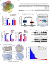
Fig. 1. MiR-375 level in PCa.
a Representative images of RNAscope technology for miR-375 in paracancerous and cancer tissues. The red dots indicate the target molecules (left). Statistical analysis of the RNAscope scores (right). MiR-375 expression in PCa from the TCGA (b) and the StarBase database (c). PRAD prostate cancer.
Fig. 2
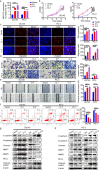
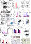
Fig. 2. MiR-375 promoted PCa cell proliferation, migration, and invasion but restrained apoptosis.
a MiR-375 overexpression in DU145 and PC-3 cells was verified via qRT-PCR after these cells were stably transfected with pSUPER-RETRO-Puro-miR-375 recombinant vector or pSUPER-RETRO-Puro-empty vector (serving as a control). CCK-8 and EdU incorporation assays were performed to assess the cell proliferation (b) and DNA synthesis rate (c) in miR-375-overexpressing DU145 and PC-3 cells. Transwell (d) and wound healing assays (e) were performed to confirm the effects of miR-375 overexpression on cell migration and invasion. f Flow cytometric analysis of the apoptotic rate in DU145 and PC-3 cells with or without miR-375 overexpression. g, h The protein levels of EMT and apoptosis markers in miR-375-overexpressing or miR-375-knockdown DU145 and PC-3 cells compared with the respective control cells. Cl. 1: Cell colony 1 transfected with pSUPER-RETRO-Puro-miR-375 recombinant vector. Cl. 2: Cell colony 2 transfected with pSUPER-RETRO-Puro-miR-375 recombinant vector. Vector: cell colony transfected with pSUPER-RETRO-Puro-empty vector (served as control). sp Cl.1: cell colony 1 transfected with pHB-U6-MCS-PGK-PURO-miR-375 sponge vector. Sp Cl.2: cell colony 2 transfected with pHB-U6-MCS-PGK-PURO-miR-375 sponge vector. NC: cell colony transfected with pHB-U6-MCS-PGK-PURO-empty vector (served as control).
Fig. 3
Fig. 3. MiR-375 targeted and negatively regulated PTPN4 in PCa.
a Potential target genes were predicted using the miRsystem, miRWalk, StarBase, TargetScan and RNA22v2 databases. A total of 50 genes were found to be shared in the five databases. b The potential binding site of miR-375 with the 3′-UTR of PTPN4 was predicted using the TargetScan database. Complementary sequences consisting of 8 nt were aligned and presented. c A luciferase assay was conducted to validate the direct binding and modulating function of miR-375 to the 3′-UTR of PTPN4. PCDH-empty: HEK-293 cells transfected with the pCDH-CMV vector. PCDH-375: HEK-293 cells transfected with pCDH-CMV vector that was subcloned with a fragment of pri-miR-375. PTPN4 WT: HEK-293 cells cotransfected with the P-MIR-Report firefly luciferase vector inserted with a fragment of the wild-type 3’-UTR of PTPN4 mRNA. PTPN4 MUT: HEK-293 cells cotransfected with the P-MIR-Report firefly luciferase vector subcloned with the 3′-UTR of PTPN4 mRNA mutated at the complementary site. d The expression of PTPN4 in PCa tissues compared with normal prostate tissues from the UALCAN database. e The correlation between miR-375 level and PTPN4 in PCa, statistically processed by StarBase. r = −0.178, p = 6.77e-5. PTPN4 at the mRNA level (f) and protein level (g), as determined by qRT-PCR and western blot in DU145 and PC-3 cells after miR-375 was overexpressed or knocked down. h IHC illustrating the differential expression of PTPN4 between 30 PCa and 17 normal tissues. The results show a representative IHC image (left) and quantitative analysis calculated by Image-Pro Plus version 6.0 software based on the 47 IHCs (right). AOD: average integrated optical density.
Fig. 4
Fig. 4. MiR-375 overexpression promotes proliferation, invasion, and migration and inhibits apoptosis via the PTPN4-STAT3 pathway.
a qRT-PCR was performed to measure the mRNA expression of PTPN4 post PTPN4 and/or miR-375 overexpression. b CCK-8 and c EdU incorporation assays were conducted to test the proliferation of DU145 and PC-3 cells in response to overexpression of miR-375 and/or PTPN4. d Transwell and e wound-healing assays were performed to determine the migration and invasion of DU145 and PC-3 cells in response to overexpression of miR-375 and/or PTPN4. f Apoptosis analysis of DU145 and PC-3 cells in response to miR-375 and/or PTPN4 overexpression. g Western blotting was conducted to evaluate p-STAT3, PTPN4, EMT- and apoptosis-related markers of DU145 and PC-3 cells in response to overexpression of miR-375 and/or PTPN4. h GSEA using the data deposited in the TCGA database suggested that PTPN4 negatively regulated the JAK-STAT pathway. NC: cells stably transfected with pSUPER-RETRO-Puro-empty vector and transiently transfected with pcDNA3.1-empty vector. PTPN4: cells stably transfected with pSUPER-RETRO-Puro-empty vector and transiently transfected with pcDNA3.1-PTPN4 recombinant vector. MiR-375: cells stably transfected with pSUPER-RETRO-Puro-miR-375 recombinant vector and transiently transfected with pcDNA3.1-empty vector. MiR-375 + PTPN4: cells stably transfected with pSUPER-RETRO-Puro-miR-375 recombinant vector and transiently transfected with pcDNA3.1-PTPN4 vector.
Fig. 5
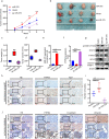
Fig. 5. MiR-375 overexpression promoted tumor growth and metastasis in vivo.
a Tumor volume during follow-up for 4 weeks. **p < 0.01, ***p < 0.001, ****p < 0.0001 vs. vector group. b Representative images of tumors in nude mice at the end timepoint. c Final tumor weight, d final tumor volume, e the expression of miR-375, and f mRNA level of PTPN4 were determined by qRT-PCR in tumor tissues at the endpoint of follow-up. g Western blotting was performed to determine the expression of p-STAT3, PTPN4, BCL2 and cleaved-PARP, and h IHC analysis of the expression of PTPN4 and Ki67 in DU145 tumor tissues as the expression of miR-375 was manipulated. i Quantification of the metastatic nodules calculated from the discontinuous lung sections. j HE staining and IHC analysis of the expression of PTPN4, E-cadherin and Vimentin using metastatic lung tissue. Vector: DU145 cells stably transfected with pSUPER-RETRO-Puro-empty vector and pHB-U6-MCS-PGK-PURO-empty vector. MiR-375: DU145 cells stably transfected with pSUPER-RETRO-Puro-miR-375 vector and pHB-U6-MCS-PGK-PURO-empty vector. Sp miR-375: DU145 cells stably transfected with pHB-U6-MCS-PGK-PURO-miR-375 sponge vector and pSUPER-RETRO-Puro-empty vector.
Fig. 6
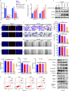
Fig. 6. E-375i inhibited PCa proliferation, migration, and invasion while promoting apoptosis in DU145 cells.
qRT-PCR illustrating miR-375 AMO loaded and intercellular transferred by hucMSC derived exosomes inhibited expression of miR-375 in DU145 and PC-3 cells (a), diminished the inhibitory effect on PTPN4 (b) in a dose-dependent manner determined by qRT-PCR, as well as downregulated the protein expression of p-STAT3 and upregulated PTPN4 (c) in a dose-dependent manner in DU145 cells, which were determined by western blot. d EdU incorporation and e CCK-8 assays were used to assess the role of miR-375 AMO-loaded exosomes on the proliferation of DU145 cells. f Transwell and g wound-healing assays were applied to test the effects of miR-375 AMO on cell migration and invasion in DU145 cells. h Flow cytometry was conducted to test the function of miR-375 AMO on the apoptotic rate of DU145 cells. i The effect of miR-375 AMO on the protein expression of p-STAT3, EMT and apoptosis markers in DU145 cells. PBS: cells treated with PBS that served as suspension control of exosomes. NC: scramble oligonucleotide. exo: exosomes suspended in PBS. e-NC: exosomes loaded with scramble oligonucleotides. e-375i: exosomes loaded with miR-375 AMO.
Fig. 7
Fig. 7. E-375i inhibited PCa proliferation in vivo.
a The flow chart showing the study design. b The bodyweight of the animals and c the tumor volume during the follow-up. d Representative images of tumors, e tumor weight, and f tumor volume at the endpoint of observation. g The expression of miR-375 and h mRNA level of PTPN4 were tested by qRT-PCR in endpoint tumor tissues. i Western blot showing the expression of p-STAT3, PTPN4, BCL2 and cleaved-PARP1 in tumor tissues. j IHC analysis of the expression of PTPN4 and Ki67 in the two groups.
Fig. 8
Fig. 8. MiR-375 regulated the expression of AR by targeting the PTPN4-STAT3 signaling pathway.
AR protein (a) and mRNA (b) levels in BPH and PCa cell lines. c The protein levels of AR in miR-375-overexpressing or miR-375-knockdown DU145 and PC-3 cells compared with the control cells. Western blotting was conducted to evaluate AR expression in DU145 and PC-3 cells in response to d overexpression of miR-375 and/or PTPN4 and e suppression of miR-375 and/or PTPN4. f The effect of miR-375 AMO on the protein expression of AR in DU145 and PC-3 cells. The mRNA expression of AR in DU145 and PC-3 cells was verified via qRT-PCR after miR-375 was overexpressed (g) or knocked down (h). i The correlation between miR-375 levels and AR in PCa, statistically processed by StarBase. r = 0.096, p = 3.28e-2. j The expression of miR-375 and k AR in PCa tissues compared with paired normal prostate tissues from TCGA database. The mRNA levels of AR in PTPN4- and/or miR-375-overexpressing (l) and PTPN4- and/or miR-375-suppressed (m) DU145 and PC-3 cells, determined by qRT-PCR. n qRT-PCR results showing the transcription levels of AR in DU145 and PC-3 cells after treatment with e-375i or the controls. o Western blot showing the expression of AR in DU145 tumor tissues as the expression of miR-375 was manipulated. p IHC analysis of AR expression in DU145 tumor tissues as miR-375 expression was manipulated. q Western blotting showing the expression of AR in tumor tissues treated with e-375i. r IHC analysis of the expression of AR in the e-375i and e-NC groups. Scramble: cells stably transfected with pHB-U6-MCS-PGK-PURO-empty vector and transiently transfected with siNC. SiPTPN4: cells stably transfected with pHB-U6-MCS-PGK-PURO-empty vector and transiently transfected with siPTPN4 vector. Sp miR-375: cells stably transfected with pHB-U6-MCS-PGK-PURO-miR-375 sponge and transiently transfected with siNC. Sp miR-375 + siPTPN4: cells stably transfected with pHB-U6-MCS-PGK-PURO-miR-375 recombinant vector and transiently transfected with siPTPN4.
Fig. 9
Fig. 9. MiR-375 regulated enzalutamide resistance by targeting the PTPN4-STAT3 signaling pathway in vitro and in vivo.
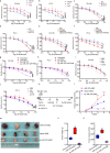
CCK-8 assays were conducted to test enzalutamide resistance of DU145 and PC-3 cells in response to a, b stably upregulated expression of miR-375, c, d stable knockdown with miR-375 sponge, e, f overexpression of miR-375 and/or PTPN4, g, h suppression of miR-375 and/or PTPN4, and i, j the use of miR-e-375i. k A flow chart showing the in vivo experimental design. l Tumor volume during follow-up for 4 weeks. m Representative images of tumors in nude mice. n Final tumor weight, o final tumor volume. ENZ: enzalutamide.
Fig. 10
Fig. 10. Schematic of e-375i elicited anti-PCa effects.
Shown is the graphical illustration of the identified pathway of miR-375 in regulating PCa progression and enzalutamide resistance.

Similar articles

Cited by

References

    1. Sung H, et al. Global cancer statistics 2020: GLOBOCAN estimates of incidence and mortality worldwide for 36 cancers in 185 countries. CA Cancer J. Clin. 2021;71:209–249. - PubMed
    1. Han Q, et al. Targeted inhibition of SIRT6 via engineered exosomes impairs tumorigenesis and metastasis in prostate cancer. Theranostics. 2021;11:6526–6541. doi: 10.7150/thno.53886. - DOI - PMC - PubMed
    1. Han Y, et al. Triptolide inhibits the AR signaling pathway to suppress the proliferation of enzalutamide resistant prostate cancer cells. Theranostics. 2017;7:1914–1927. doi: 10.7150/thno.17852. - DOI - PMC - PubMed
    1. Wang Y, et al. Mechanisms of enzalutamide resistance in castration-resistant prostate cancer and therapeutic strategies to overcome it. Br. J. Pharmacol. 2021;178:239–261. doi: 10.1111/bph.15300. - DOI - PubMed
    1. Mollaei H, Safaralizadeh R, Rostami Z. MicroRNA replacement therapy in cancer. J. Cell Physiol. 2019;234:12369–12384. doi: 10.1002/jcp.28058. - DOI - PubMed

Publication types

MeSH terms

Substances