. 2022 Aug 30;12(1):14791.
doi: 10.1038/s41598-022-19231-3.

Localized efficacy of environmental RNAi in Tetranychus urticae

Affiliations

Localized efficacy of environmental RNAi in Tetranychus urticae

Nicolas Bensoussan et al. Sci Rep. .

Abstract

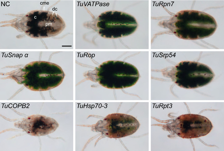
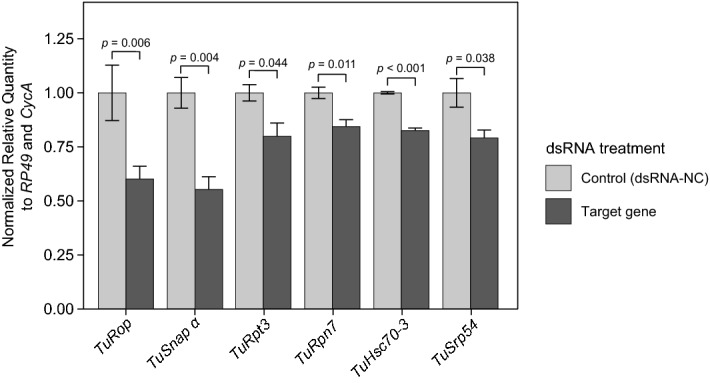
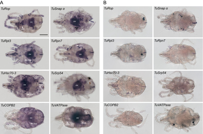
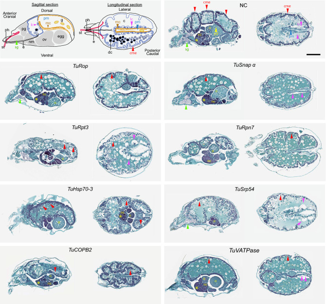
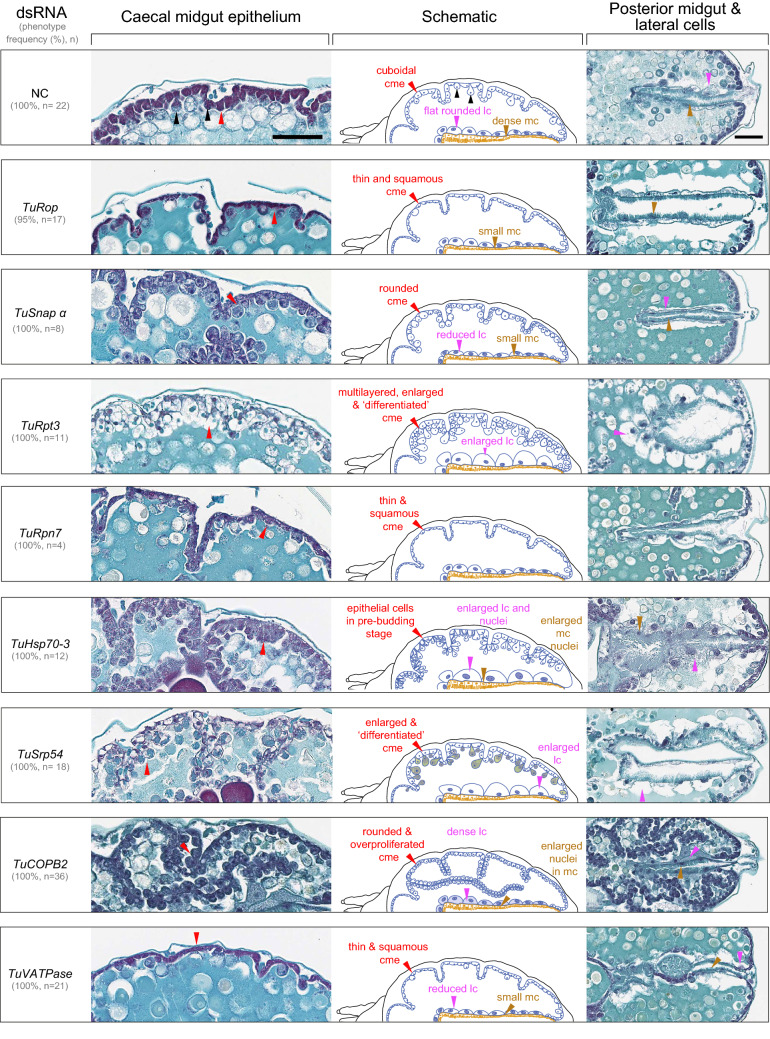
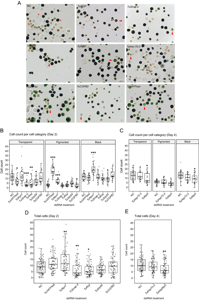
Environmental RNAi has been developed as a tool for reverse genetics studies and is an emerging pest control strategy. The ability of environmental RNAi to efficiently down-regulate the expression of endogenous gene targets assumes efficient uptake of dsRNA and its processing. In addition, its efficiency can be augmented by the systemic spread of RNAi signals. Environmental RNAi is now a well-established tool for the manipulation of gene expression in the chelicerate acari, including the two-spotted spider mite, Tetranychus urticae. Here, we focused on eight single and ubiquitously-expressed genes encoding proteins with essential cellular functions. Application of dsRNAs that specifically target these genes led to whole mite body phenotypes-dark or spotless. These phenotypes were associated with a significant reduction of target gene expression, ranging from 20 to 50%, when assessed at the whole mite level. Histological analysis of mites treated with orally-delivered dsRNAs was used to investigate the spatial range of the effectiveness of environmental RNAi. Although macroscopic changes led to two groups of body phenotypes, silencing of target genes was associated with the distinct cellular phenotypes. We show that regardless of the target gene tested, cells that displayed histological changes were those that are in direct contact with the dsRNA-containing gut lumen, suggesting that the greatest efficiency of the orally-delivered dsRNAs is localized to gut tissues in T. urticae.


Conflict of interest statement

The authors declare no competing interests.

Figures

Figure 1
Figure 1
Mite body phenotypes upon dsRNA treatment. Two-spotted spider mites, T. urticae, are characterised by the presence of two dark spots, see NC, when observed under bright field. Spots are formed by the accumulation of dark digestive cells (dc) at the anterior of the midgut caeca (c) that can be seen through the semi-transparent cutile. Mites treated with dsRNAs targeting TuVATPase, TuRpn7, TuSnap α, TuRop and TuSrp54 have a dark body phenotype characterized by the accumulation of a dark green pigment in the caeca lumen. Mites treated with dsRNAs targeting TuCOPB2, TuHsc70-3 and TuRpt3 displayed a spotless phenotype, characterized by the absence of the accumulation of black digestive cells. caeca (c); caecal midgut epithelium (cme); posterior midgut (pm); digestive cells (dc). Scale bar: 100 μm.
Figure 2
Figure 2
Target gene knockdown after dsRNA treatment. Average gene expression level relative to the expression of the reference control genes RP49 and CycA. Data represent the mean ± SE. The RT-qPCR analysis was assessed at the whole mite level and was conducted in three independent experimental runs. Statistical analysis was performed using unpaired two-tailed t test (exact P-values corresponding to each pairwise comparison between the control and the treatment are displayed).
Figure 3
Figure 3
Localization of the ingested fluorescently labelled dsRNAs-TuCOPB2-100 and dsRNAs-TuCOPB2-400 following 24 h of mite feeding. (A) Fluorescence of labeled dsRNAs is seen in the mite caeca lumen (c) and digestive cells (dc); free fluorescein-12-UTP localizes in the posterior midgut (pm); guanine pellets (g), located in the posterior midgut, exhibit autofluorescence. (B) A close-up of fluorescently labelled dsRNAs in digestive cells, posterior midgut, and epidermis. caecal midgut epithelium (cme); large cell (lc); epidermis (ep). Scale bar: (A) 100 μm; (B) 10 μm for the digestive cells panels and 25 μm for the posterior midgut and epidermis panels.
Figure 4
Figure 4
Whole-mount in situ hybridization of TuRop, TuSnap α, TuRpt3, TuRpn7, TuHsc70-3, TuSrp54, TuCOPB2 and TuVATPase in adult T. urticae females. Anti-sense (A) and sense (B) digoxigenin-labeled probes. Description of the expression domains are presented in Supplemental Table 3. caecal midgut epithelium (cme); digestive cells (dc); posterior midgut (pm); nervous mass (nm); ovaries (ov); epithelium (ep). Scale bar: 100 μm.
Figure 5
Figure 5
Histological analysis of mite RNAi phenotypes. Schematics depicting the internal anatomy of T. urticae female (top left, adapted from Bensoussan et al.). On the left side of the panel—sagittal, and on the right—longitudinal sections through mite body. Stylet (st); pharynx (ph); esophagus (e); caeca (c); digestive cell (dc); microvilli cells in posterior midgut (mc in pm); prosomal gland (pg); nervous mass (nm); guanine pellet (g). Tissues of interest are labeled with arrowheads: red, caecal midgut epitheli cells (cme); magenta, large cells (lc); blue, posterior midgut (pm); green, silk gland (sg); yellow, ovaries: asterisk, previtellogenic cells; nc nurse cells; o oocyte; yd yolk droplets within the oocyte. Higher magnifications of tissues with perturbed cellular morphologies are shown in Fig. 6. Scale bar: 100 μm.
Figure 6
Figure 6
Histological phenotypes of midgut epithelial cells upon dsRNA treatments. Left, details of caecal midgut epithelial cells; right, details of large and posterior midgut cells in mites treated with dsRNAs; middle, schematics illustrating changes of midgut epithelial cells upon dsRNA treatments. Caecal midgut epithelial cells normally consist of cuboidal and densely stained cells (red arrowhead), rounded cells and enlarged cells that protrude into the caecal lumen (black arrowheads). magenta arrowhead, large cells (lc); brown arrowhead, microvilli cells in the posterior midgut. Scale bar: 50 μm.
Figure 7
Figure 7
The effect of dsRNA treatments on digestive cells. (A) Representative images of digestive cells dissected from RNAi-treated mites. Digestive cells undergo maturation from transparent, pigmented to black, see cells labeled with asterisk in NC panel. (B,C) Cell count per cell type category: transparent, pigmented, and black at 2 days (B) and 4 days (C) post RNAi treatment. (D,E) Total cell counts at 2 days (D) and 4 days (E) post RNAi treatment. The box indicates the first and the third quartile, the middle line represents the median, and the whiskers indicate the 1.5 interquartile range. Individual data points are plotted as grey circles. Statistical analysis was performed using a negative binomial regression followed by posthoc pairwise comparisons between estimated marginal means of each RNAi treatment against the control group with subsequent P-value adjustment following the Bonferroni method (not significant, P > 0.05; *P < 0.05; **P < 0.01; ***P < 0.001).

Similar articles

Cited by

References

    1. Whangbo JS, Hunter CP. Environmental RNA interference. Trends Genet. 2008;24:297–305. - PubMed
    1. Fire A, et al. Potent and specific genetic interference by double-stranded RNA in Caenorhabditis elegans. Nature. 1998;391:806–811. - PubMed
    1. Fraser AG, et al. Functional genomic analysis of C. elegans chromosome I by systematic RNA interference. Nature. 2000;408:325–330. - PubMed
    1. Gönczy P, et al. Functional genomic analysis of cell division in C. elegans using RNAi of genes on chromosome III. Nature. 2000;408:331–336. - PubMed
    1. Ulrich J, et al. Large scale RNAi screen in Tribolium reveals novel target genes for pest control and the proteasome as prime target. BMC Genom. 2015;16:1–9. - PMC - PubMed

Publication types

Substances