. 2022 Aug 30;5(1):887.
doi: 10.1038/s42003-022-03845-4.

Homodimerized cytoplasmic domain of PD-L1 regulates its complex glycosylation in living cells

Affiliations

Homodimerized cytoplasmic domain of PD-L1 regulates its complex glycosylation in living cells

Li Zhou et al. Commun Biol. .

Abstract

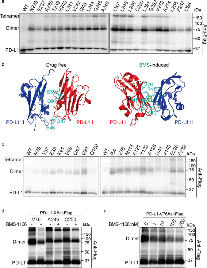
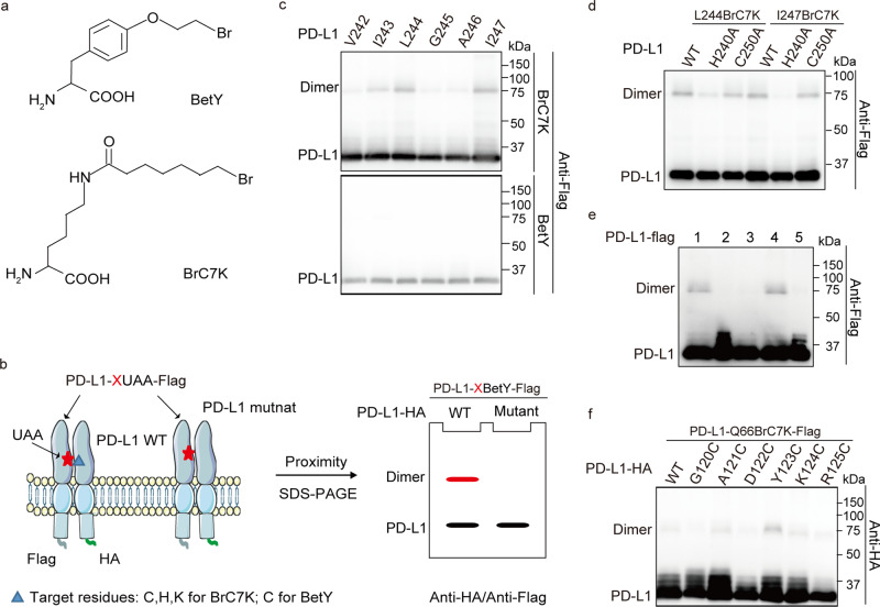
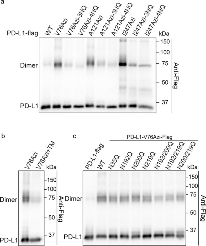
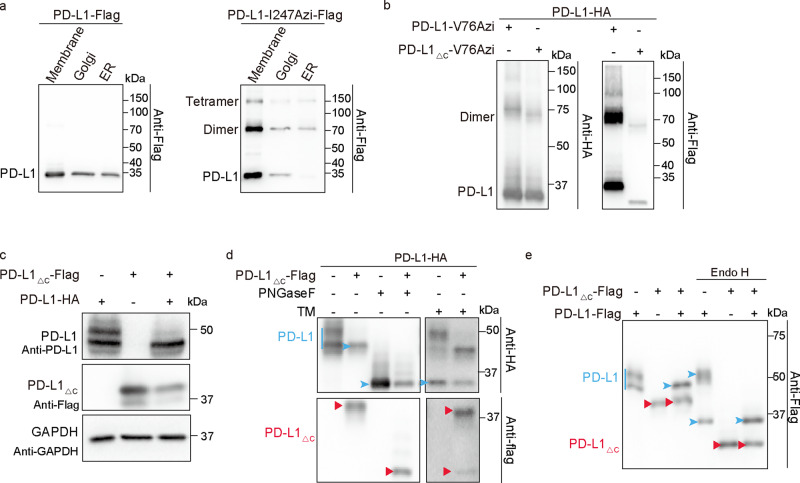
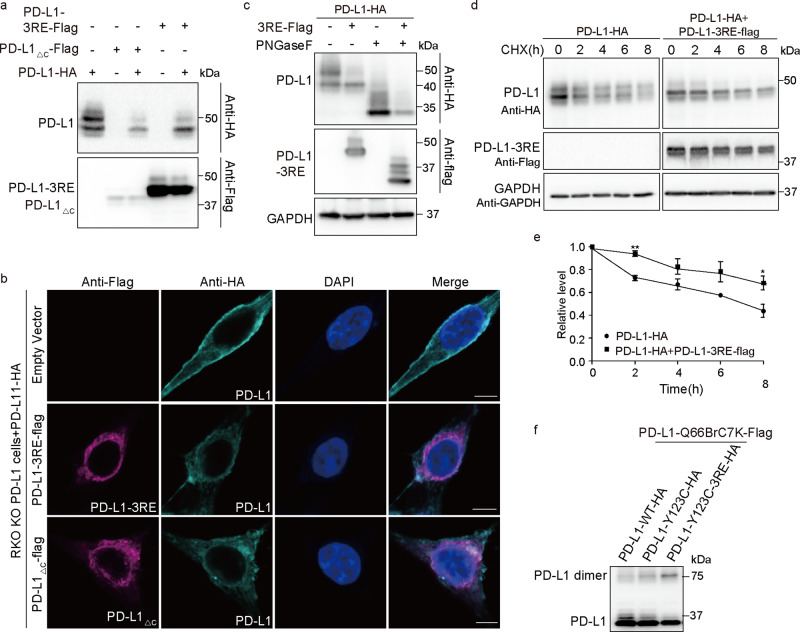
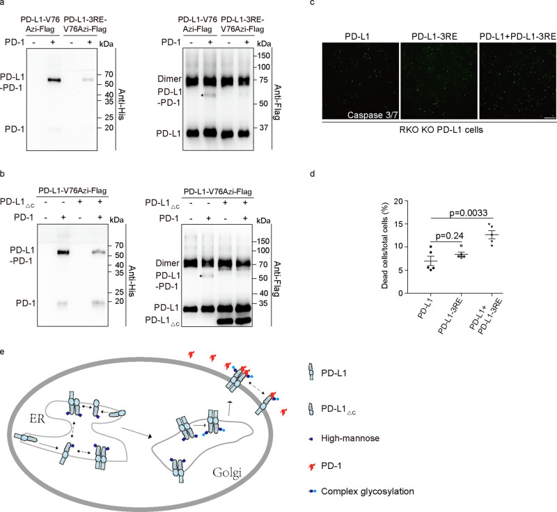
Whether membrane-anchored PD-L1 homodimerizes in living cells is controversial. The biological significance of the homodimer waits to be expeditiously explored. However, characterization of the membrane-anchored full-length PD-L1 homodimer is challenging, and unconventional approaches are needed. By using genetically incorporated crosslinkers, we showed that full length PD-L1 forms homodimers and tetramers in living cells. Importantly, the homodimerized intracellular domains of PD-L1 play critical roles in its complex glycosylation. Further analysis identified three key arginine residues in the intracellular domain of PD-L1 as the regulating unit. In the PD-L1/PD-L1-3RE homodimer, mutations result in a decrease in the membrane abundance and an increase in the Golgi of wild-type PD-L1. Notably, PD-1 binding to abnormally glycosylated PD-L1 on cancer cells was attenuated, and subsequent T-cell induced toxicity increased. Collectively, our study demonstrated that PD-L1 indeed forms homodimers in cells, and the homodimers play important roles in PD-L1 complex glycosylation and T-cell mediated toxicity.


Conflict of interest statement

The authors declare no competing interests.

Figures

Fig. 1
Fig. 1. PD-L1 homodimerized in living cells.
a Schematic diagram of genetically incorporated photocross-linking UAAs covalent capture PD-L1 homodimer. UAAs incorporated positions were referred with X or Y. PD-L1-X/YAzi-Flag, and WT PD-L1-HA were coexpressed in living cells. The anti-HA positive cross-linking band (colored red) is the complex of PD-L1-HA captured by PD-L1-XAzi-Flag. Red stars indicated the incorporated UAAs. Green curves indicated the HA tag and light blue curves indicated the Flag tag. b Structural formula of the 4-Azido-l-phenylalanine (Azi). c, d PD-L1-I247Azi captures homodimer dependent on the UAA incorporation and UV treatment. c Cells expressing PD-L1-I247Azi capture cross-linking bands in correspondence to PD-L1 homodimer. HEK293T cells were transiently transfected with PD-L1-I247tag-Flag and pIRE4-Azi plasmids. 1 mM of Azi was added to the medium. Cell lysates were separated on SDS-PAGE gels. Immunoblotting was performed using anti-Flag antibodies. d HEK293T cells were cotransfected with PD-L1-I247tag-Flag, pIRE4-Azi and PD-L1-HA plasmids. The quantity of PD-L1-I247tag-Flag plasmid increased from lane 4 to lane 6. All samples were treated with PNGase F to remove the N-glycan.
Fig. 2
Fig. 2. The transmembrane domain and extracellular domain of PD-L1 are involved in homodimerization.
a Transmembrane domain of PD-L1 is involved in its homodimerization. Residues that were replaced by Azi are indicated in the upper row. WT PD-L1 treated with UV was used as a control. HEK293T cells were irradiated with UV before being lysed. Shown are immunoblotting using anti-Flag antibodies. b Structure of drug-free and BMS-induced PD-L1 homodimer. The PDB ID is 3FN3 for drug-free PD-L1 homodimer and 5N2F for BMS-induced PD-L1 homodimer. BMS was colored green. Residues that are important for PD-L1 homodimerization were colored cyan. c Screening of residues in extracellular domain that covalent captured PD-L1 homodimer. Transfected cells expressing PD-L1-XAzi-Flag proteins were treated with UV to induce cross-linking and performed immunoblotting analysis. d, e BMS-1166 reduces Azi-mediated covalent capture of PD-L1 homodimerization dosage dependently. HEK293T cells were transiently transfected with PD-L1-Xtag-Flag and pIRE4-Azi plasmids. d Residues replaced by Azi are indicated in the upper row. 100 nM of BMS-1166 were incubated with cells for 18 h before UV treatment. e Working concentration of BMS-1166 ranged from 0 to 200 nM. All samples were treated with PNGase F to remove the N-glycan.
Fig. 3
Fig. 3. PD-L1 asymmetrically homodimerized.
a Structural formula of proximity-enabled UAAs, BrC7K, and BetY. b Schematic diagram of mapping interacting residue pairs using the chemical cross-linking UAAs. Red stars represent the proximity-enabled UAAs, namely BrC7K/BetY. BrC7K/BetY selectively reacts with targeted amino acids (blue triangle) to form covalent complexes which are destroyed by targeted amino acids mutation. c BrC7K incorporated into the transmembrane domain of PD-L1 capture its homodimers. PD-L1-Xtag-Flag plasmid was cotransfected with pHY-XYRS or pHY-BrC7KRS to incorporate the BetY or BrC7K individually. Samples were collected for anti-Flag immunoblotting analysis. Residues replaced by BrC7K/BetY are indicated in the upper row. d BrC7K at L244 and I247 of PD-L1 interact with H240 residues. PD-L1-L244/I247BrC7K-Flag with or without the H240A/C250A mutation were expressed in HEK293T cells. e D284 in the intracellular domain of PD-L1 interacts with C272. BrC7K were incorporated into D284 of PD-L1 or PD-L1 mutants. Lane 1: PD-L1-D284BrC7K-Flag; Lane 2: PD-L1-C272A-Flag; Lane 3: PD-L1-C272A-D284BrC7K-Flag; Lane 4: PD-L1-D284BrC7K-H286A-Flag; Lane 5: PD-L1-Flag. f Q66 and Y123 in the extracellular domain of PD-L1 are paired interaction residues. Residues mutated to Cys are indicated in the upper row. All samples were treated with PNGase F to remove the N-glycan.
Fig. 4
Fig. 4. Glycosylation plays an important role in PD-L1 homodimerization.
a, b Defect of PD-L1 glycosylation result in PD-L1 homodimerization attenuated. a PD-L1 and PD-L1-3NQ/4NQ mutants were incorporated with Azi. Azi introduced positions are indicated in the upper row. Cells were treated with UV for 20 min before collecting. b Tunicamycin (TM) suppresses PD-L1 homodimerization. Cells expressing PD-L1-V76Azi were treated with 5 µM tunicamycin for 24 h before collection. c The number of glycosylated residues affects homodimerization of PD-L1. Azi was incorporated into V76 of WT PD-L1-Flag or PD-L1-Flag mutants. All samples were treated with PNGase F to remove the N-glycan.
Fig. 5
Fig. 5. Homodimerized intracellular domains regulate PD-L1 complex glycosylation.
a PD-L1 forms more homodimers in the Endoplasmic reticulum. HEK293T cells were transiently transfected with PD-L1-I247tag-Flag and pIRE4-Azi plasmids. 1 mM of Azi was added to the medium. Cells were treated with UV for 15 min. The ER, Golgi, and plasma membrane fractionation were isolated and separated on SDS-PAGE gels. Immunoblotting was performed using anti-Flag antibodies. b PD-L1△C homodimerized with WT PD-L1. Azi was genetically incorporated into V76 of PD-L1-Flag or PD-L1△C-Flag in HEK293T cells expressing PD-L1-HA. Cells were treated with UV for 20 min. Cell lysates were treated with PNGase F to remove the N-glycan. c, e PD-L1△C suppresses complex glycosylation of WT PD-L1. c, d WT PD-L1-HA and PD-L1△C-Flag plasmids were transfected into RKO KO PD-L1 Cells. c PD-L1△C affects the pattern of PD-L1 that showed on immunoblotting. The anti-PD-L1 antibodies (clone number E1L3N) could recognize the full-length PD-L1, but not the PD-L1△C. d Cell lysates for lane 3 and lane 4 were treated with PNGase F. Samples for lane 5 and lane 6 were treated with 10 µM tunicamycin (TM) for 24 h before cell collecting. e RKO KO PD-L1 cells were cotransfected with PD-L1-Flag and PD-L1△C-Flag plasmids. Cell lysates for lanes 4–6 were treated with Endo H. Lines and arrowheads colored blue indicate full-length PD-L1. Red arrowheads indicate the PD-L1△C proteins.
Fig. 6
Fig. 6. Dimerized key arginine residues in the intracellular domain play a critical role in PD-L1 complex glycosylation.
a PD-L1-3RE mutant suppresses complex glycosylation of WT PD-L1. PD-L1-HA plasmid was cotransfected with PD-L1-3RE-Flag or PD-L1△C-Flag into RKO KO PD-L1 cells. b Plasma membrane localization of WT PD-L1 attenuates in cells expressing PD-L1-3RE mutant. Shown are immunofluorescence of cells expressing PD-L1-HA and PD-L1-3RE-Flag/PD-L1△C-Flag. Representative images are shown for each condition. Scale bars, 5 μm. c Total expression level of WT PD-L1 decreased in cells expressing PD-L1-3RE. RKO KO PD-L1 cells were transfected with PD-L1-HA or cotransfected with PD-L1-HA and PD-L1-3RE-Flag. Samples of lanes 3–4 were de-glycosylated with PNGase F. d, e PD-L1-3RE stabilized highly mannosylated PD-L1. PD-L1-HA or PD-L1-HA and PD-L1-3RE-Flag were transfected into RKO KO PD-L1 cells. The duration that cells treated with 150 µM CHX was labeled at the upper row. The quantification data represent the mean ± SEM for three independent experiments. *P ≤ 0.05, **P ≤ 0.01. f Effect of 3RE mutation on conformation of the PD-L1 ECD homodimer. Samples were treated with PNGase F to remove the N-glycan.
Fig. 7
Fig. 7. PD-L1 homodimerization plays an important role in PD-1 binding and T-cell toxicity.
a PD-L1-Azi interaction with PD-1 was suppressed by 3RE mutation. Azi was incorporated at V76 of WT PD-L1 or PD-L1-3RE mutant. HEK293T cells were irradiated with UV in the absence or presence of PD-1-His protein. Shown are immunoblotting using anti-Flag and anti-His antibodies. b PD-L1△C suppressed WT PD-L1 cross-linking with PD-1. Cells expressing PD-L1-V76Azi-Flag or coexpressing PD-L1-V76Azi-Flag and PD-L1△C-Flag were incubated with PD-1-His protein. Covalent cross-linking was induced by UV treatment for 20 min. c, d T-cell mediated tumor cell killing assay in RKO KO PD-L1 cells expressing PD-L1 and PD-L1-3RE. Green fluorescent was counted as dead cells. Representative phases are shown. Scale bar, 100 μm. d The ratio of dead cells was quantified. N = 5. e Schematic diagram of PD-L1 homodimer function. All samples were treated with PNGase F to remove the N-glycan.

Similar articles

Cited by

References

    1. Ahmadzadeh M, et al. Tumor antigen-specific CD8 T cells infiltrating the tumor express high levels of PD-1 and are functionally impaired. Blood. 2009;114:1537–1544. doi: 10.1182/blood-2008-12-195792. - DOI - PMC - PubMed
    1. Herbst RS, et al. Predictive correlates of response to the anti-PD-L1 antibody MPDL3280A in cancer patients. Nature. 2014;515:563–567. doi: 10.1038/nature14011. - DOI - PMC - PubMed
    1. Sharma P, Allison JP. The future of immune checkpoint therapy. Science. 2015;348:56–61. doi: 10.1126/science.aaa8172. - DOI - PubMed
    1. Boussiotis VA. Molecular and biochemical aspects of the PD-1 checkpoint pathway. N. Engl. J. Med. 2016;375:1767–1778. doi: 10.1056/NEJMra1514296. - DOI - PMC - PubMed
    1. Robert C, et al. Nivolumab in previously untreated melanoma without BRAF mutation. N. Engl. J. Med. 2015;372:320–330. doi: 10.1056/NEJMoa1412082. - DOI - PubMed

Publication types