. 2022 Aug 30;12(1):14799.
doi: 10.1038/s41598-022-19239-9.

Porphyromonas gingivalis-mediated disruption in spiral artery remodeling is associated with altered uterine NK cell populations and dysregulated IL-18 and Htra1

Affiliations

Porphyromonas gingivalis-mediated disruption in spiral artery remodeling is associated with altered uterine NK cell populations and dysregulated IL-18 and Htra1

Tanvi Tavarna et al. Sci Rep. .

Abstract

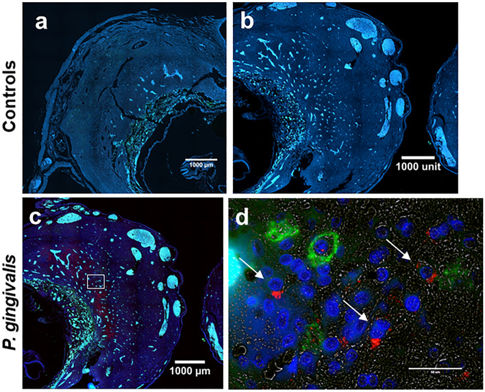
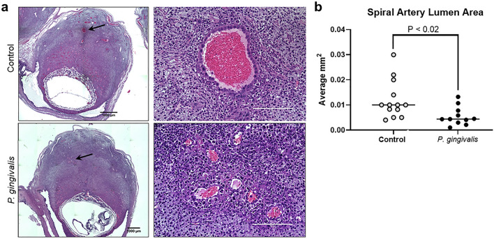
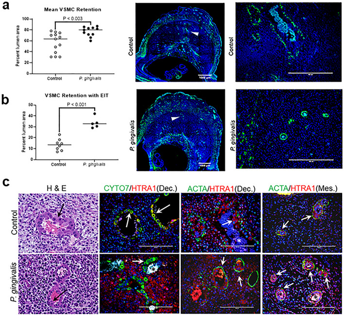
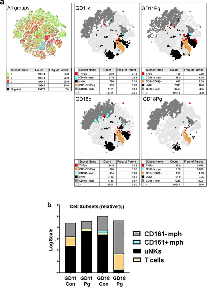
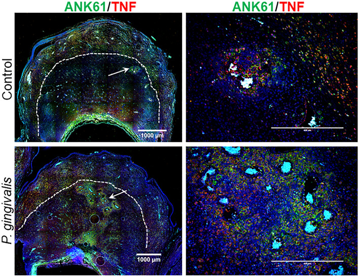
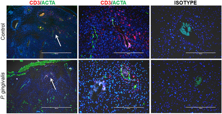
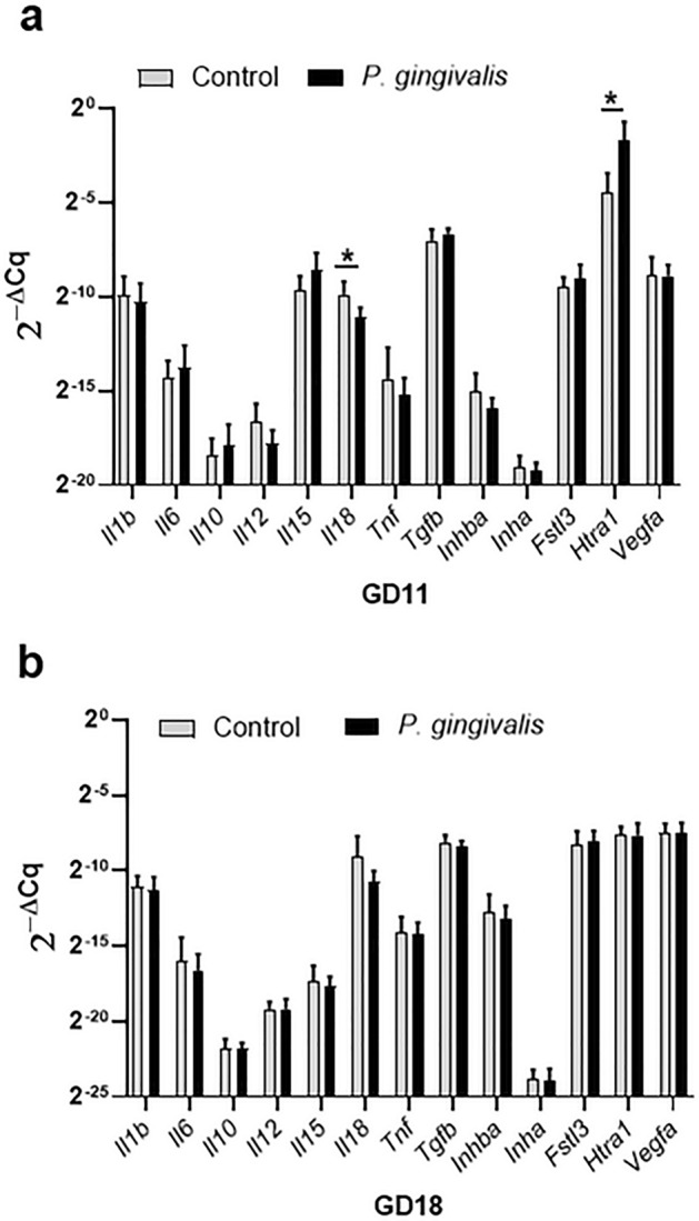
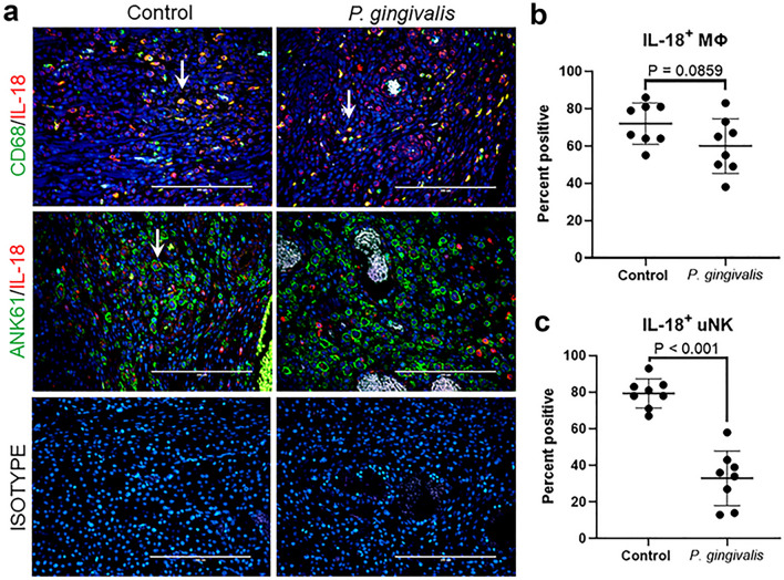
Impaired spiral artery remodeling (IRSA) underpins the great obstetrical syndromes. We previously demonstrated that intrauterine infection with the periodontal pathogen, Porphyromonas gingivalis, induces IRSA in rats. Since our previous studies only examined the end stage of arterial remodeling, the aim of this study was to identify the impact of P. gingivalis infection on the earlier stages of remodeling. Gestation day (GD) 11 specimens, a transition point between trophoblast-independent remodeling and the start of extravillous trophoblast invasion, were compared to late stage GD18 tissues. P. gingivalis was found in decidual stroma of GD11 specimens that already had reduced spiral artery remodeling defined as smaller arterial lumen size, increased retention of vascular smooth muscle, and decreased invasion by extravillous trophoblasts. At GD11, P. gingivalis-induced IRSA coincided with altered uterine natural killer (uNK) cell populations, decreased placental bed expression of interleukin-18 (IL-18) with increased production of temperature requirement A1 (Htra1), a marker of oxidative stress. By GD18, placental bed IL-18 and Htra1 levels, and uNK cell numbers were equivalent in control and infected groups. However, infected GD18 placental bed specimens had decreased TNF + T cells. These results suggest disturbances in placental bed decidual stroma and uNK cells are involved in P. gingivalis-mediated IRSA.


Conflict of interest statement

The authors declare no competing interests.

Figures

Figure 1
Figure 1
Representative images of P. gingivalis in GD11 specimens. (a) Stained section from an uninfected control. (b) Corresponding isotype control for P. gingivalis positive specimen. (c) Low magnified image of a P. gingivalis positive specimen. Boxed region indicates the region that is pictured in (d). Transillumination (grey) was used to define the tissue architecture in decidual stroma in (d). Scale bar = 50 µm. (a–c) Are tiled composites of individual pictures taken at × 10 magnification with an EVOS Auto FL imaging system. Scale bar = 1000 µm. Tissue sections were dual stained with an antibody to NK cell marker, a-natural killer cell activation structures (green) and P. gingivalis (red, white arrows). DAPI (blue) was used as a nuclear stain.
Figure 2
Figure 2
Impact of P. gingivalis infection on GD11 spiral artery lumen size. (a) Representative images from control and P. gingivalis-infected specimens. Left panels are tiled composites of individual pictures taken at × 10 magnification with an EVOS Auto FL imaging system. Scale bar = 1000 µm. Black arrows mark the spiral artery segments pictured in the corresponding right panels. White scale bar = 200 µm. (b) Spiral artery lumen area measurements from control and P. gingivalis-infected specimens. Horizontal lines indicate the mean ± SD. Data was analyzed by student’s t test.
Figure 3
Figure 3
Retention of spiral arterial VSMC and invasive endovascular trophoblasts in GD11 specimens. (a) Mean percent circumferential area of spiral arterial VSMCs in specimens with and without endovascular invasive trophoblast (EIT). Each data point represents the average value of all area measurements taken within the middle portion of the utero-placenta. Horizontal bars are mean ± SD. Representative images of spiral arterial VSMC detected by staining for smooth muscle actin (ACTA, green) and DAPI staining for nuclei. Left panels are tiled composites of individual pictures taken at × 10 magnification with an EVOS Auto FL imaging system. Scale bar = 1000 µm. White arrowheads mark the spiral artery segments pictured in the corresponding right panels. White scale bar = 200 µm. (b) Mean percent circumferential area of VSMC in spiral arterial loops containing EIT. Horizontal lines = mean ± SD. (c) Representative images of EIT in hematoxylin and eosin stained sections (H&E), and by immunostaining with an antibody to cytokeratin 7(CYTO7, green) and high temperature requirement A1 (Htra1, red). Graphical data was analyzed by student’s t test.
Figure 4
Figure 4
Overview of placental bed leukocyte populations identified by flow cytometry. (a) Clustering analysis by tSNE demonstrated population differences free of batch effects and identified corresponding immune cell populations in each treatment group. Processed datasets from all treatments (n = 4 per group) were analyzed with FlowJo’s t-SNE and Flow Means plug-in (FlowJo v10.6.2, Treestar, Ashland, OR). (b) Bar chart shows relative proportion of each immune cell subset included in the analysis. Individual populations are shown in supplement file, Fig. S3b.
Figure 5
Figure 5
In situ distribution of uNK cells in control and P. gingivalis-infected specimens collected at GD11. uNK cells were identified with an antibody to Ank61 (green), some of which are also positive for TNF (red). Left panels are tiled composites of individual pictures taken at × 10 magnification with an EVOS Auto FL imaging system. Scale bar = 1000 µm. Dashed lines demarcate the junction between decidua and myometrium. White arrows mark the spiral artery segments pictured in corresponding magnified images on the right (white scale bar = 200 µm). Representative images of GD18 specimens and isotype controls are found in Supplement File Fig. S5.
Figure 6
Figure 6
In situ detection of T cells (CD3+, red) and VSMC (ACTA+, green) in GD18 placental bed sections. Left panels are low magnification (× 4) images, scale bar = 1000 µm. White arrows mark the spiral artery segment pictured in the corresponding right panels. White scale bar = 200 µm. All images were captured with an EVOS Auto FL imaging system.
Figure 7
Figure 7
Placental bed gene expression profiles of GD11 (a) and GD18 (b) specimens. Cytokine and chemokine gene expression measured by RT-qPCR. Values are the mean 2−ΔCq ± SD determined by the comparative Cq method using Actb as the reference gene (n = 10). *Indicates 2−ΔCq values that were significantly different (P < 0.05) by unpaired student’s t test.
Figure 8
Figure 8
Impact of P. gingivalis infection on IL-18 (red) positive MΦ (CD68, green) and uNK cells (Ank61, green) in GD11 specimens. (a) Representative images of CD68+/IL-18+ MΦs or Ank61+/IL-18+ uNK cells taken with an EVOS Auto FL imaging system. White arrows indicate double positive cells. Scale bar = 200 µm. (b) Percent CD68+/IL-18+ MΦs. (c) Percent Ank61+/IL-18+ uNK cells. Horizontal lines in each graph indicate the mean ± SD. Data was analyzed by unpaired student’s t test.

Similar articles

Cited by

References

    1. Brosens I, Pijnenborg R, Vercruysse L, Romero R. The, "Great Obstetrical Syndromes" are associated with disorders of deep placentation. Am. J. Obstet. Gynecol. 2011;204:193–201. doi: 10.1016/j.ajog.2010.08.009. - DOI - PMC - PubMed
    1. Almasry SM, Elmansy RA, Elfayomy AK, Algaidi SA. Ultrastructure alteration of decidual natural killer cells in women with unexplained recurrent miscarriage: A possible association with impaired decidual vascular remodelling. J. Mol. Histol. 2015;46:67–78. doi: 10.1007/s10735-014-9598-8. - DOI - PubMed
    1. El-Azzamy H, et al. Dysregulated uterine natural killer cells and vascular remodeling in women with recurrent pregnancy losses. Am. J. Reprod. Immunol. 2018;80:e13024. doi: 10.1111/aji.13024. - DOI - PubMed
    1. Leon R, et al. Detection of Porphyromonas gingivalis in the amniotic fluid in pregnant women with a diagnosis of threatened premature labor. J. Periodontol. 2007;78:1249–1255. doi: 10.1902/jop.2007.060368. - DOI - PubMed
    1. Barak S, Oettinger-Barak O, Machtei EE, Sprecher H, Ohel G. Evidence of periopathogenic microorganisms in placentas of women with preeclampsia. J. Periodontol. 2007;78:670–676. doi: 10.1902/jop.2007.060362. - DOI - PubMed

Publication types

Substances