. 2022 Jul 26;4(1):vdac116.
doi: 10.1093/noajnl/vdac116. eCollection 2022 Jan-Dec.

Real-time PACS-integrated longitudinal brain metastasis tracking tool provides comprehensive assessment of treatment response to radiosurgery

Affiliations

Real-time PACS-integrated longitudinal brain metastasis tracking tool provides comprehensive assessment of treatment response to radiosurgery

Gabriel Cassinelli Petersen et al. Neurooncol Adv. .

Abstract

Background: Treatment of brain metastases can be tailored to individual lesions with treatments such as stereotactic radiosurgery. Accurate surveillance of lesions is a prerequisite but challenging in patients with multiple lesions and prior imaging studies, in a process that is laborious and time consuming. We aimed to longitudinally track several lesions using a PACS-integrated lesion tracking tool (LTT) to evaluate the efficiency of a PACS-integrated lesion tracking workflow, and characterize the prevalence of heterogenous response (HeR) to treatment after Gamma Knife (GK).

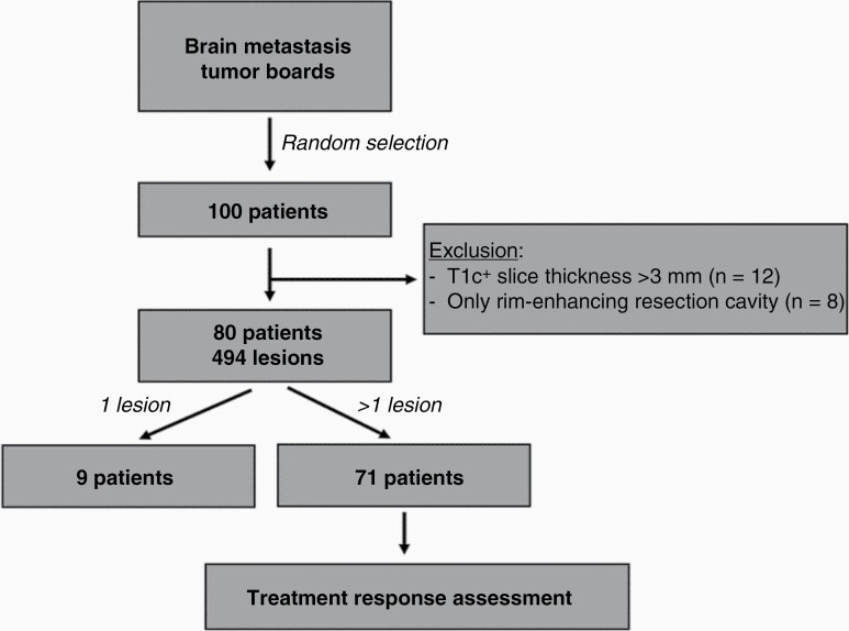
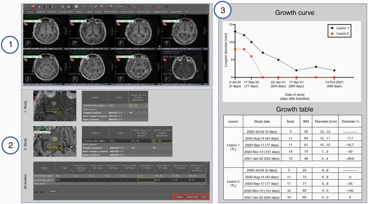
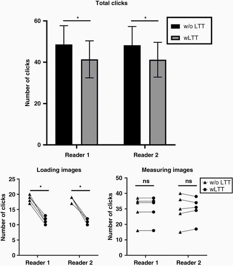
Methods: We selected a group of brain metastases patients treated with GK at our institution. We used a PACS-integrated LTT to track the treatment response of each lesion after first GK intervention to maximally seven diagnostic follow-up scans. We evaluated the efficiency of this tool by comparing the number of clicks necessary to complete this task with and without the tool and examined the prevalence of HeR in treatment.

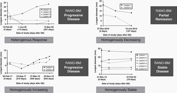
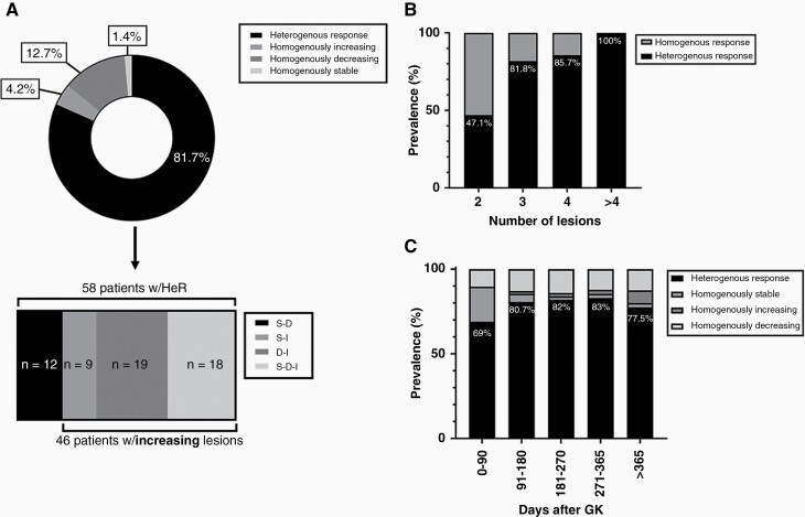
Results: A cohort of eighty patients was selected and 494 lesions were measured and tracked longitudinally for a mean follow-up time of 374 days after first GK. Use of LTT significantly decreased number of necessary clicks. 81.7% of patients had HeR to treatment at the end of follow-up. The prevalence increased with increasing number of lesions.

Conclusions: Lesions in a single patient often differ in their response to treatment, highlighting the importance of individual lesion size assessments for further treatment planning. PACS-integrated lesion tracking enables efficient lesion surveillance workflow and specific and objective result reports to treating clinicians.

Keywords: brain metastasis tracking; heterogenous response; lesion; radiosurgery; response to treatment.


Figures

Figure 1.
Figure 1.
Patient selection workflow. From the randomly selected 100 patients, 80 were included in the analysis and measured longitudinally. From this group, nine had only one lesion and homogeneity of treatment response could be assessed in seventy-one patients.
Figure 2.
Figure 2.
Workflow of the lesion tracking tool (LTT) for longitudinal tracking of metastases. Step 1: Loading of the images and deployment of the hanging protocol. Step 2: Individual lesions are measured over time and the results recorded and automatically assigned to their corresponding study date. Step 3: The output of the LTT is generated. The automatically generated treatment response curves and tables allow for qualitative and quantitative assessment of lesion growth. In this example, two metastatic lesions are tracked for 408 days after Gamma Knife (GK) radiosurgery over seven follow-ups post GK, revealing that both lesions remitted homogenously after treatment.
Figure 3.
Figure 3.
Comparison of clicks necessary to measure and track lesions with and without the LTT. Mean total click count was significantly lower with use of LTT (Reviewer 1: 48.6 vs 41.4 clicks, P < .001; Reviewer 2: 48.2 vs 41.2 clicks, P < .001). Similarly, mean click count for loading the images was significantly lower when the LTT was used (Reviewer 1 and 2: 11.4 vs 18.6 clicks, P < .001). There was no significant difference between the number of clicks necessary for the size measurement of the lesions itself (Reviewer 1: 30 vs 30 clicks; Reviewer 2: 29.8 vs 29.6 clicks, P = .84). * = P < .001, ns, not significant.
Figure 4.
Figure 4.
Types of treatment response after GK therapy demonstrating growth curves with homogenous and heterogenous treatment responses in comparison to treatment response calculated by RANO-BM. 3A and 3B demonstrate progressive disease as classified by RANO-BM criteria, but when looking at growth curves, 3A demonstrates heterogenous response with only one lesion needing treatment. On the other hand, 3B demonstrates homogeneously increasing growth curves, thus there is a need to treat both lesions in this patient. 3C demonstrates partial remission with homogeneous decrease in size of all lesions over time. 3D demonstrates stable disease on both RANO-BM and growth curve assessment. These treatment response curves show that while an overall assessment of treatment response is necessary, in some cases it is also advantageous to look at the change in size of individual lesions: while the overall sum of diameters is increasing in Lesion 1 and slightly in Lesion 3, Lesions 2 and 4 have both remitted or are remained stable after treatment.
Figure 5.
Figure 5.
Distribution of lesions according to their response types: (A) Prevalence of different treatment responses at last date of follow-up among patients with >1 lesion. The lower graph dissects the group of heterogenous response (HeR), into the different components that make up HeR. S-D: stable and decreasing, S-I: stable and increasing, D-I: decreasing and increasing, S-D-I: stable, decreasing, and increasing lesions. (B) The chart indicates the prevalence of HeR in relation to homogenous response as a function of the total number of lesions per patient. (C) The graph shows the proportion of different treatment responses on follow-up scans as a function of the number of days after first GK intervention.

References

    1. Nayak L, Lee EQ, Wen PY. Epidemiology of brain metastases. Curr Oncol Rep. 2012;14(1):48–54. - PubMed
    1. Chen JY, Vedantham S, Lexa FJ. Burnout and work-work imbalance in radiology-wicked problems on a global scale. A baseline pre-COVID-19 survey of US neuroradiologists compared to international radiologists and adjacent staff. Eur J Radiol. 2022:110153. doi:10.1016/j.ejrad.2022.110153. - DOI - PubMed
    1. Benson J, Burgstahler M, Zhang L, Rischall M. The value of structured radiology reports to categorize intracranial metastases following radiation therapy. Neuroradiol J. 2019;32(4):267–272. - PMC - PubMed
    1. Bink A, Benner J, Reinhardt J, et al. . Structured reporting in neuroradiology: intracranial tumors. Front Neurol. 2018;9:32. - PMC - PubMed
    1. Crusz SM, Tang YZ, Sarker SJ, et al. . Heterogeneous response and progression patterns reveal phenotypic heterogeneity of tyrosine kinase inhibitor response in metastatic renal cell carcinoma. BMC Med. 2016;14(1):185. - PMC - PubMed

LinkOut - more resources