

Application of CT Imaging in Differential Diagnosis and Nursing of Endocrine Tumors
- PMID: 36043145
- PMCID: PMC9377953
- DOI: 10.1155/2022/4071081
Application of CT Imaging in Differential Diagnosis and Nursing of Endocrine Tumors
Retraction in
-
Retracted: Application of CT Imaging in Differential Diagnosis and Nursing of Endocrine Tumors.Contrast Media Mol Imaging. 2023 Oct 4;2023:9831473. doi: 10.1155/2023/9831473. eCollection 2023. Contrast Media Mol Imaging. 2023. PMID: 37829317 Free PMC article.
Abstract
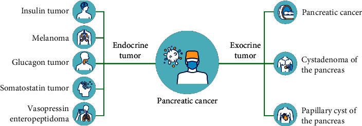
In order to investigate the value of preoperative X-ray computed tomography (CT) in predicting the pathological grade of pancreatic neuroendocrine tumors. This paper retrospectively analyzed the CT image examination of pancreatic neuroendocrine tumors, the image characteristics of G-NEC detected by CT image, and the detection of GST by spiral CT. In order to clearly diagnose and evaluate the size and scope of the focus, whether there is adjacent tissue invasion, metastasis, and treatment effect, CT, MR, PET-CT, nuclide specific imaging, and other imaging methods are widely used in the medical treatment of pNEN patients. These imaging methods have the advantages of noninvasive, rapid imaging, objective image medium, and strong repeatability. If the pathological grade of pNEN patients can be obtained by imaging examination before operation, it will be of great benefit to the formulation of treatment strategies and the prediction of clinical outcomes. Combining CT image performance with imaging omics characteristics to establish a prediction model that can develop a better auxiliary decision-making tool for clinical practice. Different pathological grades prompt clinicians to provide personalized and accurate medical treatment for patients, and reduce excessive medical treatment or wrong judgment caused by unclear preoperative diagnostic information.
Copyright © 2022 Xue Jiang et al.
Conflict of interest statement
The authors declare that they have no conflicts of interest.
Figures
References
-
- Zhang Z. C., Hu J., Kong Y. Y., Ren M., Cai X. Application of immunohistochemical staining of bcl-2, ber-ep4, cd10, ck20, and ki-67 in differential diagnosis between trichoblastoma and basal cell carcinoma. Zhonghua bing li xue za zhi Chinese journal of pathology . 2021;50(4):376–381. doi: 10.3760/cma.j.cn112151-20200722-00587. - DOI - PubMed
Publication types
MeSH terms
LinkOut - more resources
Full Text Sources
Medical
Research Materials
