. 2023 Feb;12(4):4951-4967.
doi: 10.1002/cam4.5187. Epub 2022 Aug 31.

Co-amplified with PDGFRA, IGFBP7 is a prognostic biomarker correlated with the immune infiltrations of glioma

Affiliations

Co-amplified with PDGFRA, IGFBP7 is a prognostic biomarker correlated with the immune infiltrations of glioma

Haiwei Wang et al. Cancer Med. 2023 Feb.

Abstract

Background: A subgroup of glioma carry genetic 4q12 amplification including platelet derived growth factor receptor α (PDGFRA) and insulin like growth factor binding protein 7 (IGFBP7). However, the prognosis of PDGFRA and IGFBP7 in glioma is unclear.

Methods: The prognosis of PDGFRA and IGFBP7 was determined using cox regression and Kaplan-Meier survival analysis. Pathways associated with IGFBP7 were analyzed through gene set enrichment analysis (GSEA). Immune profiling of glioma was determined using "ESTIMATE" and "TIMER" database.

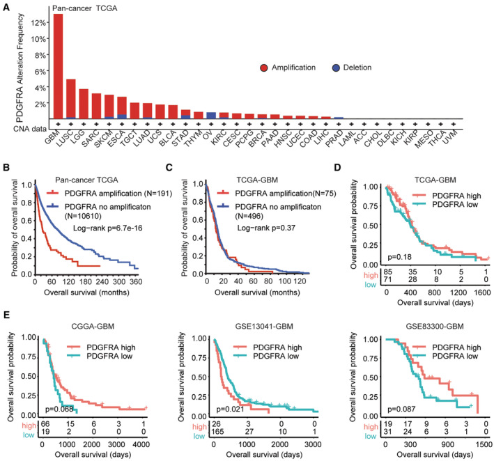
Results: PDGFRA amplification or expression was not correlated with the outcomes of glioblastoma (GBM). IGFBP7 but not PDGFRA was over-expressed in GBM. IGFBP7 over-expression was correlated with the unfavorable outcomes of GBM. In lower grade glioma (LGG), PDGFRA over-expression was not correlated with the unfavorable prognosis of LGG, while, IGFBP7 was a prognostic biomarker of LGG. LGG patients with IGFBP7 lower expressions had prolonged clinical overall survival. Combination of IDH mutation, LGG grade and IGFBP7 achieved even better prognostic effects in LGG. Moreover, IGFBP7 was over-expressed in glioma patients with wild type IDH or with high grades. IGFBP7 over-expression was correlated with the unfavorable outcomes of glioma. Furthermore, IGFBP7 was hypo-methylated in GBM or LGG patients without IDH mutations. IGFBP7 hyper-methylation was correlated with the lower overall survival of GBM or LGG. LGG patients with wild type IDH and with IGFBP7 hypo-methylation demonstrated even worse prognosis. IGFBP7 was associated with multiple immune-related signaling pathways in GBM or LGG. The stromal score, immune score and the infiltrations of immune cells were also correlated with IGFBP7 and the prognosis of LGG.

Conclusions: IGFBP7 but not PDGFRA served an ideal prognostic marker and therapeutic target of glioma.

Keywords: IGFBP7; PDGFRA; glioblastoma; immune infiltrations; lower grade glioma.


Conflict of interest statement

The authors declare that they have no conflict of interest.

Figures

FIGURE 1
FIGURE 1
Prognostic relevance of PDGFRA amplification and expression. (A) Copy number alterations of PDGFRA in pan‐cancer TCGA datasets. (B) Overall survival of tumor patients without or with PDGFRA amplifications in pan‐cancer TCGA datasets. (C) Overall survival of GBM patients without or with PDGFRA amplifications in TCGA‐GBM dataset. (D) Overall survival of GBM patients with PDGFRA higher expressions or with PDGFRA lower expressions in TCGA‐GBM dataset. (E) Correlations of overall survival and PDGFRA expression were validated in CGGA‐GBM, GSE13041‐GBM and GSE83300‐GBM datasets
FIGURE 2
FIGURE 2
Expression and prognosis of PDGFRA co‐amplified genes. (A) Co‐occurrence of PDGFRA, KIT, KDR, CHIC2, EXOC1, IGFBP7, RASL11B, and USP46 amplification in TCGA‐GBM. (B) PDGFRA, KIT, KDR, CHIC2, EXOC1, IGFBP7, RASL11B and USP46 expressions in normal brain tissues and TCGA‐GBM tissues. (C) PDGFRA, KIT, KDR, CHIC2, EXOC1, IGFBP7, RASL11B and USP46 expression levels in normal brain tissues and GSE68848‐GBM tissues. (D) Associations of PDGFRA, KIT, KDR, CHIC2, EXOC1, IGFBP7, RASL11B and USP46 expression with GBM overall survival in TCGA‐GBM, CGGA‐GBM GSE13041‐GBM and GSE83300‐GBM datasets. p value and HR were determined through cox regression
FIGURE 3
FIGURE 3
IGFBP7 over‐expression is correlated with the progression of GBM. (A) Correlations of IGFBP7 with GBM overall survival in TCGA‐GBM, CGGA‐GBM, GSE13041‐GBM and GSE83300‐GBM datasets were demonstrated through The Kaplan–Meier plots. (B) IGFBP7 expressions in GBM mesenchymal, classical, proneural and neural subtypes of TCGA‐GBM and GSE13041‐GBM. (C) IGFBP7 expressions in IDH mutant GBM patients or IDH non‐mutant GBM patients in TCGA‐GBM and CGGA‐GBM datasets. (D) Correlations of age of diagnosis, gender, IDH mutation and IGFBP7 expression in the predication of the GBM overall survival in TCGA‐GBM and CGGA‐GBM datasets. p value and HR were determined by multivariate cox regression
FIGURE 4
FIGURE 4
IGFBP7 over‐expression is correlated with the progression of LGG. (A) Oncoprint of IDH1 mutation, PDGFRA amplification and IGFBP7 amplification in TCGA‐LGG dataset. (B) Overall survival of LGG patients without or with PDGFRA amplifications in TCGA‐LGG dataset. (C) Forest plots revealed the associations of PDGFRA expression with LGG overall survival in TCGA‐LGG, CGGA‐LGG and GSE107850‐LGG datasets. (D) Overall survival of LGG patients with PDGFRA higher expressions or with PDGFRA lower expressions in TCGA‐LGG, CGGA‐LGG and GSE107850‐LGG datasets. (E) Forest plots revealed the associations of IGFBP7 expression with LGG overall survival in TCGA‐LGG, CGGA‐LGG and GSE107850‐LGG datasets. (F) Overall survival of LGG patients with IGFBP7 higher expressions or with IGFBP7 lower expressions in TCGA‐LGG, CGGA‐LGG and GSE107850‐LGG datasets
FIGURE 5
FIGURE 5
Independent prognostic effects of IGFBP7 in LGG. (A) Correlations of age of glioma diagnosis, gender, glioma grade, IDH mutation and IGFBP7 expression with LGG clinical overall survival in TCGA‐LGG and CGGA‐LGG datasets were determined by multivariate cox regression. (B) IGFBP7 expression levels in IDH mutant LGG patients or IDH wild type LGG patients in TCGA‐LGG and CGGA‐LGG datasets. (C) Associations of IGFBP7 with clinical overall survival of IDH wild type LGG patients in TCGA‐LGG and CGGA‐LGG datasets. (D) Associations of IGFBP7 with clinical overall survival of IDH mutant LGG patients in TCGA‐LGG and CGGA‐LGG datasets. (E) Overall survival of grade 2 IGFBP7 highly expressed LGG patients, grade 3 IGFBP7 highly expressed LGG patients, grade 2 IGFBP7 lowly expressed LGG patients and grade 3 IGFBP7 lowly expressed LGG patients in TCGA‐LGG and CGGA‐LGG datasets
FIGURE 6
FIGURE 6
Expression and prognosis of IGFBP7 in glioma. (A) IGFBP7 expression levels in grade 2, 3 and 4 glioma patients. (B) IGFBP7 expression levels in GBM and LGG patients in GSE4412 and GSE43378 datasets. (C) Associations of IGFBP7 and overall survival of glioma in TCGA‐GBMLGG, CGGA‐GBMLGG, GSE4412‐GBMLGG and GSE43378‐GBMLGG. (D) Associations of IGFBP7 and overall survival of glioma in TCGA‐GBMLGG, CGGA‐GBMLGG, GSE4412‐GBMLGG and GSE43378‐GBMLGG datasets
FIGURE 7
FIGURE 7
IGFBP7 hypo‐methylation is correlated with the progression of glioma. (A) IGFBP7 methylation levels in LGG or GBM patients without or with IDH mutations. (B) Methylation levels of IGFBP7 in grade 2, 3 or 4 glioma patients. (C) Clinical outcomes of GBM patients with IGFBP7 hyper‐methylation or with IGFBP7 hypo‐methylation in TCGA‐GBM and CGGA‐GBM datasets. (D) Overalls survival of IGFBP7 hyper‐methylated LGG patients and IGFBP7 hypo‐methylated LGG patients in TCGA‐LGG and CGGA‐LGG datasets. (E) Overalls survival of IGFBP7 hypo‐methylated IDH wild type LGG patients and IGFBP7 hyper‐methylated IDH wild type LGG patients in TCGA‐LGG and CGGA‐LGG datasets. (F) Overalls survival of IGFBP7 hypo‐methylated glioma patients and IGFBP7 hyper‐methylated glioma patients in TCGA‐GBMLGG and CGGA‐GBMLGG datasets
FIGURE 8
FIGURE 8
Identification of genes associated with IGFBP7. (A) Volcano plots showed the changed genes in IGFBP7 highly expressed GBM patients in TCGA‐GBM and CGGA‐GBM datasets. (B) Overlapped differentially expressed genes in IGFBP7 highly expressed GBM patients in TCGA‐GBM and CGGA‐GBM datasets. (C) Volcano plots showed the differentially expressed genes in IGFBP7 highly expressed LGG patients in TCGA‐LGG and CGGA‐LGG datasets. (D) Overlapped differentially expressed genes in IGFBP7 highly expressed LGG patients in TCGA‐LGG and CGGA‐LGG datasets. (E) Signaling pathways associated with IGFBP7 in GBM patients were determined by DAVID online tools. (F) Signaling pathways associated with IGFBP7 in LGG patients. (G) Transcription factors associated with IGFBP7 in LGG patients
FIGURE 9
FIGURE 9
Immune‐related pathways and NF‐κB transcription factor are correlated with IGFBP7. (A) Enriched signaling pathways in GBM patients with higher IGFBP7 expressions or lower IGFBP7 expressions in TCGA‐GBM and CGGA‐GBM datasets. (B) Enriched signaling pathways in LGG patients with higher IGFBP7 expressions or lower IGFBP7 expressions in TCGA‐LGG and CGGA‐LGG datasets. (C) Enrichment of NF‐κB in LGG patients with higher IGFBP7 expressions or lower IGFBP7 expressions in TCGA and CGGA datasets.
FIGURE 10
FIGURE 10
Correlations of IGFBP7 and immune infiltration of glioma. (A) Spearman correlations of IGFBP7 with stromal and immune score in TCGA‐GBM and CGGA‐GBM datasets. (B) Spearman correlations of IGFBP7 with stromal and immune score in TCGA‐LGG and CGGA‐LGG datasets. (C) Spearman correlations of IGFBP7 and the infiltrations of six immune‐related cell types in GBM patients in TCGA‐GBM dataset. (D) Prognosis of the infiltrations of six different immune cells in TCGA‐GBM dataset. (E) Spearman correlations of IGFBP7 and the infiltrations of six immune‐related cell types in TCGA‐LGG dataset. (F) Prognosis of the infiltrations of six different immune cells in TCGA‐LGG dataset

Similar articles

References

    1. Wen PY, Kesari S. Malignant gliomas in adults. N Engl J Med. 2008;359:492‐507. doi:10.1056/NEJMra0708126 - DOI - PubMed
    1. Ostrom QT, Gittleman H, Liao P, et al. CBTRUS statistical report: primary brain and central nervous system tumors diagnosed in the United States in 2007‐2011. Neuro Oncol. 2014;16(Suppl 4):iv1‐iv63. doi:10.1093/neuonc/nou223 - DOI - PMC - PubMed
    1. Jiang T, Nam DH, Ram Z, et al. Clinical practice guidelines for the management of adult diffuse gliomas. Cancer Lett. 2020;499:60‐72. doi:10.1016/j.canlet.2020.10.050 - DOI - PubMed
    1. Stupp R, Brada M, van den Bent MJ, et al. High‐grade glioma: ESMO clinical practice guidelines for diagnosis, treatment and follow‐up. Ann Oncol. 2014;25(Suppl 3):iii93‐iii101. doi:10.1093/annonc/mdu050 - DOI - PubMed
    1. Louis DN, Perry A, Reifenberger G, et al. The World Health Organization classification of tumors of the central nervous system: A summary. Acta Neuropathol. 2016;131:803‐820. doi:10.1007/s00401-016-1545-1 - DOI - PubMed

Publication types

Substances