. 2022 Sep 20;204(9):e0024822.
doi: 10.1128/jb.00248-22. Epub 2022 Aug 31.

Identification and Characterization of the Alternative σ28 Factor in Treponema denticola

Affiliations

Identification and Characterization of the Alternative σ28 Factor in Treponema denticola

Kurni Kurniyati et al. J Bacteriol. .

Abstract

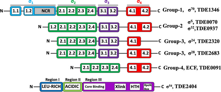
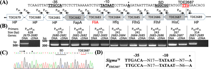
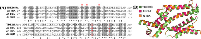
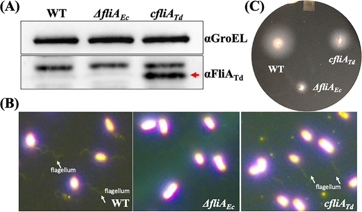
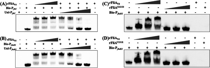
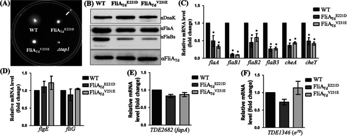
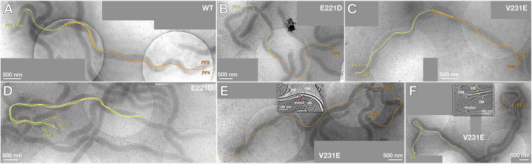
FliA (also known as σ28), a member of the bacterial σ70 family of transcription factors, directs RNA polymerase to flagellar late (class 3) promoters and initiates transcription. FliA has been studied in several bacteria, yet its role in spirochetes has not been established. In this report, we identify and functionally characterize a FliA homolog (TDE2683) in the oral spirochete Treponema denticola. Computational, genetic, and biochemical analyses demonstrated that TDE2683 has a structure similar to that of the σ28 of Escherichia coli, binds to σ28-dependent promoters, and can functionally replace the σ28 of E. coli. However, unlike its counterparts from other bacteria, TDE2683 cannot be deleted, suggesting its essential role in the survival of T. denticola. In vitro site-directed mutagenesis revealed that E221 and V231, two conserved residues in the σ4 region of σ28, are indispensable for the binding activity of TDE2683 to the σ28-dependent promoter. We then mutated these two residues in T. denticola and found that the mutations impair the expression of flagellin and chemotaxis genes and bacterial motility as well. Cryo-electron tomography analysis further revealed that the mutations disrupt the flagellar symmetry (i.e., number and placement) of T. denticola. Collectively, these results indicate that TDE2683 is a σ28 transcription factor that regulates the class 3 gene expression and controls the flagellar symmetry of T. denticola. To the best of our knowledge, this is the first report establishing the functionality of FliA in spirochetes. IMPORTANCE Spirochetes are a group of medically important but understudied bacteria. One of the unique aspects of spirochetes is that they have periplasmic flagella (PF, also known as endoflagella) which give rise to their unique spiral shape and distinct swimming behaviors and play a critical role in the pathophysiology of spirochetes. PF are structurally similar to external flagella, but the underpinning mechanism that regulates PF biosynthesis and assembly remains largely unknown. By using the oral spirochete Treponema denticola as a model, this report provides several lines of evidence that FliA, a σ28 transcriptional factor, regulates the late flagellin gene (class 3) expression, PF assembly, and flagellar symmetry as well, which provides insights into flagellar regulation and opens an avenue to investigate the role of σ28 in spirochetes.

Keywords: Treponema; flagella; motility; sigma factors; spirochetes.


Conflict of interest statement

The authors declare no conflict of interest.

Figures

FIG 1
FIG 1
Schematic illustration of σs in T. denticola. These factors are annotated in the genome of T. denticola (42). Individual domains in these σ factors were identified by BLAST (Fig. S3) and SMART searches and then aligned to their counterparts (i.e., σ70, σ28, and σ54) from E. coli and other bacteria (1, 4–6, 8, 10, 90, 91).
FIG 2
FIG 2
TDE2683 is located at a large gene cluster regulated by a σ70 promoter. (A) Diagram showing the genes adjacent to TDE2683. Arrows represent the relative positions and orientations of RT-PCR primers that span the intergenic regions between individual ORFs. (B) RT-PCR analysis. For each pair of primers, chromosomal DNA was used as a positive control. The numbers below the primers are predicted sizes of RT-PCR and PCR products. (C) 5′ RACE analysis. The arrow shows the sequencing direction, and an asterisk indicates the transcriptional start site. (D) Sequence comparison between the E. coli σ70 promoter and the promoter sequence identified upstream of TDE2687 (designated PTDE2687).
FIG 3
FIG 3
TDE2683 is a homolog of FliA. (A) Sequence alignment of FliA homologs. TDE2683 (WP_002667112) is aligned with E. coli FliA (NP_416432), S. Typhimurium FliA (NP_460909), and B. subtilis SigD (WP_187704716). Only partial alignment is shown. Asterisks represent three invariable residues in the family of FliA. The residues in red are to be mutated. The alignment was analyzed using Clustal Omega. (B) Homology modeling shows that TDE2683 (FliATd) has structural topology similar to that of the FliA proteins (σ28) of E. coli and S. Typhimurium. The figure was generated by the Swiss-Model server using FliAEc (PDB ID 1SC5) (8) as a template.
FIG 4
FIG 4
FliATd substitutes for the function of FliAEc in E. coli. (A) Detection of FliATd in E. coli by immunoblotting analysis. For this experiment, equivalent amounts (~10 μg) of whole-cell lysates of three E. coli strains were analyzed by SDS-PAGE and then probed with specific antibodies against E. coli GroEL (αGroEL) and T. denticola FliA (αFliATd). GroEL was used as a loading control. WT, E. coli DH5α; ΔfliAEc, fliA deletion mutant of E. coli; cfliATd, engineered E. coli strain in which the entire fliAEc gene was replaced in frame with fliATd. (B) Visualization of E. coli flagella. E. coli cells were first stained with Ryu as previously described (82) and then visualized under dark-field illumination at ×100 magnification using a Zeiss Axiostar Plus microscope. (C) Swimming plate assay. This assay was carried out on swimming plates containing 1% tryptone, 1% NaCl, and 0.4% Bacto agar, as previously documented (87). The plates were incubated at 37°C overnight.
FIG 5
FIG 5
EMSA shows that FliATd binds to two σ28-dependent promoters. (A) EMSA using E. coli fliC promoter (PfliC) as a DNA probe. For this assay, biotin-labeled PfliC (Bio-PfliC) was incubated with increasing amounts of wild-type FliATd recombinant protein (rFliATd) and titrated with increasing concentrations of unlabeled PfliC (Unl-PfliC). (B to D) EMSA using T. denticola flaB2 promoter (PflaB2) as a DNA probe. Biotin-labeled PflaB2 (Bio-PflaB2) was incubated with increasing amounts of rFliATd (B) or two mutated recombinant proteins, rFliAE221D (C) and rFliAV231E (D), and competed out with increasing concentrations of unlabeled PflaB2 (Unl-PflaB2).
FIG 6
FIG 6
Characterizations of two fliATd site-directed mutants of T. denticola. (A) Swimming plate assay of WT, FliATdE221D, and FliATdV231E strains. This assay was carried out on 0.35% agarose plates containing TYGVS medium diluted 1:5 with PBS. The plates were incubated anaerobically at 37°C for 5 days. A nonmotile Δtap1 mutant was used as a control to determine initial inoculum sizes. (B) Immunoblotting analysis of WT, FliATdE221D, and FliATdV231E strains. Equivalent amounts of whole-cell lysates were analyzed by SDS-PAGE and then probed with specific antibodies to DnaK, FlaA, and FlaBs. DnaK was used as a loading control. (C to F) qRT-PCR analysis of WT, FliATdE221D, and FliATdV231E strains. For this experiment, the levels of four flagellar filament genes (flaA, flaB1, flaB2, and flaB3), two chemotaxis genes (cheA and cheY), two flagellar genes (flgE and fliG), TDE2682 (fapA), and TDE134670) were measured by qRT-PCR, as previously described (33). The dnaK gene transcript was used as an internal control to normalize the readouts of qRT-PCR. The results are expressed as the level of individual gene transcripts in two mutants relative to that in WT. *, P < 0.05.
FIG 7
FIG 7
Whole-cell cryo-ET analysis of T. denticola wild type and two mutants. (A) Projection image of a WT cell; (B to F) projection images of FliATdE221D and FliATdV231E mutant cells. PF originating from one end are colored in yellow, and those from the opposite end are in orange. The boxed images in panel E and F show the detailed structure of PF and flagellar protrusions. PF, periplasmic flagella; OM, outer membrane; IM, inner membrane. Each panel was generated by combining multiple high-resolution cryo-ET images; the image contrast is different among these data.

Similar articles

Cited by

References

    1. Feklistov A, Sharon BD, Darst SA, Gross CA. 2014. Bacterial sigma factors: a historical, structural, and genomic perspective. Annu Rev Microbiol 68:357–376. 10.1146/annurev-micro-092412-155737. - DOI - PubMed
    1. Helmann JD. 2019. Where to begin? Sigma factors and the selectivity of transcription initiation in bacteria. Mol Microbiol 112:335–347. 10.1111/mmi.14309. - DOI - PMC - PubMed
    1. Saecker RM, Record MT, Jr, Dehaseth PL. 2011. Mechanism of bacterial transcription initiation: RNA polymerase-promoter binding, isomerization to initiation-competent open complexes, and initiation of RNA synthesis. J Mol Biol 412:754–771. 10.1016/j.jmb.2011.01.018. - DOI - PMC - PubMed
    1. Liu B, Hong C, Huang RK, Yu Z, Steitz TA. 2017. Structural basis of bacterial transcription activation. Science 358:947–951. 10.1126/science.aao1923. - DOI - PubMed
    1. Malhotra A, Severinova E, Darst SA. 1996. Crystal structure of a sigma 70 subunit fragment from E. coli RNA polymerase. Cell 87:127–136. 10.1016/S0092-8674(00)81329-X. - DOI - PubMed

Publication types

MeSH terms

LinkOut - more resources