Association of Artificial Intelligence-Aided Chest Radiograph Interpretation With Reader Performance and Efficiency
- PMID: 36044215
- PMCID: PMC9434361
- DOI: 10.1001/jamanetworkopen.2022.29289
Association of Artificial Intelligence-Aided Chest Radiograph Interpretation With Reader Performance and Efficiency
Abstract
Importance: The efficient and accurate interpretation of radiologic images is paramount.
Objective: To evaluate whether a deep learning-based artificial intelligence (AI) engine used concurrently can improve reader performance and efficiency in interpreting chest radiograph abnormalities.
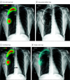
Design, setting, and participants: This multicenter cohort study was conducted from April to November 2021 and involved radiologists, including attending radiologists, thoracic radiology fellows, and residents, who independently participated in 2 observer performance test sessions. The sessions included a reading session with AI and a session without AI, in a randomized crossover manner with a 4-week washout period in between. The AI produced a heat map and the image-level probability of the presence of the referrable lesion. The data used were collected at 2 quaternary academic hospitals in Boston, Massachusetts: Beth Israel Deaconess Medical Center (The Medical Information Mart for Intensive Care Chest X-Ray [MIMIC-CXR]) and Massachusetts General Hospital (MGH).
Main outcomes and measures: The ground truths for the labels were created via consensual reading by 2 thoracic radiologists. Each reader documented their findings in a customized report template, in which the 4 target chest radiograph findings and the reader confidence of the presence of each finding was recorded. The time taken for reporting each chest radiograph was also recorded. Sensitivity, specificity, and area under the receiver operating characteristic curve (AUROC) were calculated for each target finding.
Results: A total of 6 radiologists (2 attending radiologists, 2 thoracic radiology fellows, and 2 residents) participated in the study. The study involved a total of 497 frontal chest radiographs-247 from the MIMIC-CXR data set (demographic data for patients were not available) and 250 chest radiographs from MGH (mean [SD] age, 63 [16] years; 133 men [53.2%])-from adult patients with and without 4 target findings (pneumonia, nodule, pneumothorax, and pleural effusion). The target findings were found in 351 of 497 chest radiographs. The AI was associated with higher sensitivity for all findings compared with the readers (nodule, 0.816 [95% CI, 0.732-0.882] vs 0.567 [95% CI, 0.524-0.611]; pneumonia, 0.887 [95% CI, 0.834-0.928] vs 0.673 [95% CI, 0.632-0.714]; pleural effusion, 0.872 [95% CI, 0.808-0.921] vs 0.889 [95% CI, 0.862-0.917]; pneumothorax, 0.988 [95% CI, 0.932-1.000] vs 0.792 [95% CI, 0.756-0.827]). AI-aided interpretation was associated with significantly improved reader sensitivities for all target findings, without negative impacts on the specificity. Overall, the AUROCs of readers improved for all 4 target findings, with significant improvements in detection of pneumothorax and nodule. The reporting time with AI was 10% lower than without AI (40.8 vs 36.9 seconds; difference, 3.9 seconds; 95% CI, 2.9-5.2 seconds; P < .001).
Conclusions and relevance: These findings suggest that AI-aided interpretation was associated with improved reader performance and efficiency for identifying major thoracic findings on a chest radiograph.
Conflict of interest statement
Figures


Comment in
-
Beyond the AJR: Improving Radiologist Performance and Efficiency Using Chest Radiography Artificial Intelligence.AJR Am J Roentgenol. 2023 Jul;221(1):143. doi: 10.2214/AJR.22.28768. Epub 2022 Nov 23. AJR Am J Roentgenol. 2023. PMID: 36416396 No abstract available.
References
Publication types
MeSH terms
LinkOut - more resources
Full Text Sources
Medical
Miscellaneous

