. 2022 Aug 31;14(660):eabj7465.
doi: 10.1126/scitranslmed.abj7465. Epub 2022 Aug 31.

A targetable pathway in neutrophils mitigates both arterial and venous thrombosis

Affiliations

A targetable pathway in neutrophils mitigates both arterial and venous thrombosis

Lalitha Nayak et al. Sci Transl Med. .

Abstract

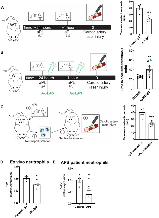
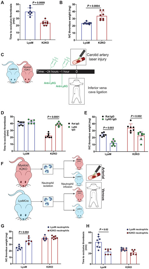
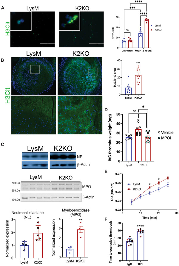
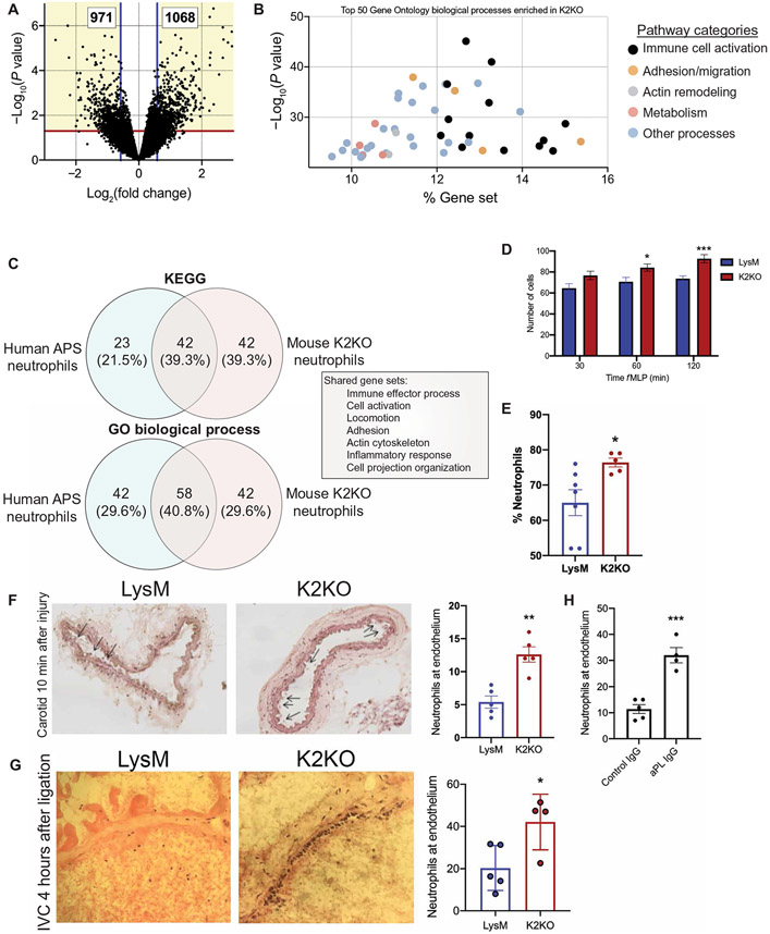
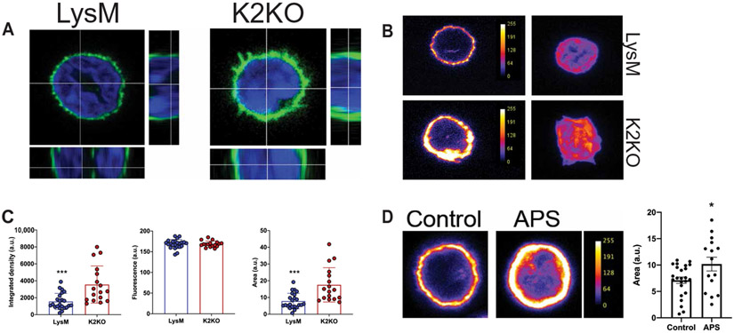
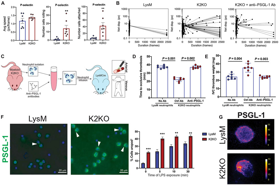
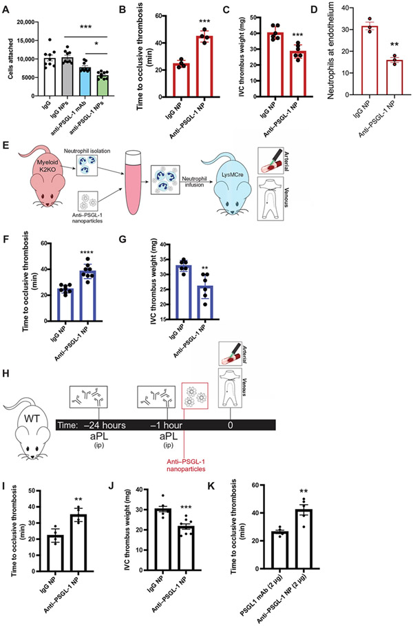
Arterial and venous thrombosis constitutes a major source of morbidity and mortality worldwide. Long considered as distinct entities, accumulating evidence indicates that arterial and venous thrombosis can occur in the same populations, suggesting that common mechanisms are likely operative. Although hyperactivation of the immune system is a common forerunner to the genesis of thrombotic events in both vascular systems, the key molecular control points remain poorly understood. Consequently, antithrombotic therapies targeting the immune system for therapeutics gain are lacking. Here, we show that neutrophils are key effectors of both arterial and venous thrombosis and can be targeted through immunoregulatory nanoparticles. Using antiphospholipid antibody syndrome (APS) as a model for arterial and venous thrombosis, we identified the transcription factor Krüppel-like factor 2 (KLF2) as a key regulator of neutrophil activation. Upon activation through genetic loss of KLF2 or administration of antiphospholipid antibodies, neutrophils clustered P-selectin glycoprotein ligand 1 (PSGL-1) by cortical actin remodeling, thereby increasing adhesion potential at sites of thrombosis. Targeting clustered PSGL-1 using nanoparticles attenuated neutrophil-mediated thrombosis in APS and KLF2 knockout models, illustrating the importance and feasibility of targeting activated neutrophils to prevent pathological thrombosis. Together, our results demonstrate a role for activated neutrophils in both arterial and venous thrombosis and identify key molecular events that serve as potential targets for therapeutics against diverse causes of immunothrombosis.


Figures

Fig. 1.
Fig. 1.. Activated neutrophils drive arterial and venous thrombosis.
(A) In the murine antiphospholipid antibody (aPL)–injected model, mice were injected intraperitoneally (ip) with either aPL isolated from patients with APS (n = 6 mice) or IgG isolated from healthy controls (n = 4 mice) at 24 hours and 1 hour before a carotid artery laser-induced injury model of thrombosis. Time to occlusive thrombus development was measured. (B) Neutrophil depletion in aPL-treated mice using anti-Ly6G antibody (n = 8 mice) or control IgG (n = 8 mice) was followed by carotid artery injury to measure dependency on neutrophils in arterial thrombus formation. Time to occlusive thrombosis development was measured. All mice received aPLs. (C) Neutrophils were harvested from IgG- or aPL-treated wild-type (WT) mice and transferred into untreated WT mice (n = 4 IgG-treated neutrophil recipients; n = 6 aPL-treated neutrophil recipients) before carotid artery injury. (D) Klf2 expression was measured in neutrophils harvested from IgG-injected (n = 3) or aPL-injected (n = 5) WT mice. (E) KLF2 expression was measured in neutrophils harvested from humans with APS (n = 7) or healthy controls (n = 4). *P < 0.05, **P < 0.01, and ***P < 0.001 by unpaired, two-tailed Student’s t test.
Fig. 2.
Fig. 2.. Neutrophil activation through loss of KLF2 worsens arterial and venous thrombosis.
(A) Time to occlusive thrombosis was measured in the carotid artery injury model comparing LysM (n = 7) and K2KO (n = 8) mice. (B) IVC thrombus weight measured after IVC ligation comparing LysM and K2KO mice. n = 8 mice. (C) The timeline shows the experimental model of neutrophil depletion studies in K2KO mice. (D and E) Neutrophils were depleted with anti-Ly6G antibody or control antibody in LysM (n = 6 to 7) and K2KO (n = 6 to 8) mice before (D) arterial or (E) venous thrombosis induction. (F) The timeline depicts adoptive transfer experiments wherein neutrophils were isolated from healthy LysM or K2KO mice and infused into LysM/K2KO mice before arterial or venous thrombosis. (G and H) Infusion of LysM (CRE) or K2KO (KO) neutrophils into respective genotypes (n = 6 to 8 per group) was followed by initiation of (G) arterial and (H) venous thrombosis. Data were analyzed using an unpaired, two-tailed Student’s t test.
Fig. 3.
Fig. 3.. Loss of KLF2 permits prothrombotic neutrophil functions.
(A) Neutrophil extracellular trap (NET) staining with citrullinated histone 3 (H3Cit) is shown for N-formyl-Met-Leu-Phe (fMLP)-stimulated LysM or K2KO neutrophils in vitro (n = 5). (B) H3Cit NET staining within harvested clots after IVC ligation was quantified (LysM, n = 9; K2KO, n = 11). Quantification represents the percentage of clot area that is H3Cit+. (C) Western blot of cell lysate from unstimulated LysM and K2KO neutrophils measuring neutrophil elastase (NE) and myeloperoxidase (MPO) abundance is shown. Quantification is normalized to β-actin. (D) LysM (n = 6) and K2KO [vehicle, n = 8; MPO inhibitor (MPOi), n = 8] mice received an MPOi before the IVC thrombosis model in mice; thrombus weight was measured. (E) A colorimetric tissue factor (TF) activity assay was used to measure conversion of factor X to Xa over time in LysM and K2KO neutrophils (n = 3 to 5). OD, optical density. (F) The effect of TF neutralization before carotid artery thrombosis was measured. K2KO neutrophils were incubated with either control or TF-neutralizing antibody (1H1) before infusion into LysM mice (IgG, n = 9; 1H1, n = 8). *P < 0.05, **P < 0.01, ***P < 0.001, and ****P < 0.0001 by unpaired, two-tailed Student’s t test.
Fig. 4.
Fig. 4.. Activated neutrophils are transcriptionally primed for prothrombotic adhesion and migration.
(A) The volcano plot from RNA sequencing of peripheral blood neutrophils demonstrates more than 2000 differentially expressed genes between K2KO and LysM cells. (B) Gene Ontology (GO) biological process analysis of differentially expressed genes from K2KO neutrophil RNA sequencing is shown. % Gene set represents the proportion of the entire process that is affected by loss of KLF2. (C) The overlap of enriched pathways between K2KO neutrophils and neutrophils from humans with APS is shown. (D) Results of a transwell migration experiment using N-formyl-Met-Leu-Phe (fMLP) as chemoattractant for LysM and K2KO neutrophils are shown (n = 10 to 12). (E) Peritoneal lavage after fMLP intraperitoneal injection was used to quantify neutrophil migration in LysM (n = 7) and K2KO (n = 5) mice. (F and G) Ly6G-stained neutrophils were quantified along vessel walls early after (F) carotid artery injury (10 min) and (G) IVC ligation (4 hours) are shown for LysM and K2KO mice (n = 4 to 5). (H) Neutrophils were quantified along vessel walls early after carotid artery injury in IgG-injected (n = 5) or aPL-injected (n = 4) WT mice. *P < 0.05, **P < 0.01, and ***P < 0.001 by unpaired, two-tailed Student’s t test.
Fig. 5.
Fig. 5.. Neutrophil activation is associated with cortical actin remodeling in K2KO and APS neutrophils.
(A) Confocal microscopy of F-actin–stained neutrophils demonstrates cortical actin alignment in untreated LysM and K2KO neutrophils. (B) Fluorescent intensity of cortical F-actin expression in untreated LysM and K2KO neutrophils is shown. (C) Quantification of actin area, fluorescence intensity, and integrated density from confocal images is shown. LysM, n = 23 cells from three mice; K2KO, n = 19 cells from three mice. (D) Cortical actin staining is shown for neutrophils isolated from either healthy controls (n = 25 cells from three donors) or patients with APS (n = 15 cells from three donors). Scales in (B) and (D) indicate cortical actin fluorescence intensity. a.u., arbitrary units. *P < 0.05 and ***P < 0.001 by unpaired, two-tailed Student’s t test.
Fig. 6.
Fig. 6.. PSGL-1 clustering facilitates prothrombotic K2KO neutrophil adhesion.
(A) Measurement of flowing speed, extent of rolling, and adhesion is shown for LysM and K2KO neutrophils in microfluidic channels coated with P-selectin. Avg, average; px, pixels. (B) Dot plots depict the duration of time that each cell remained in the field of view. Neutrophils from LysM mice, K2KO mice, and K2KO mice pretreated ex vivo with anti–PSGL-1 antibody (Ab) were compared. (C) An experimental outline is shown where neutrophils were isolated from K2KO mice, incubated with either control or anti–PSGL-1 antibodies, and infused into LysM mice before the arterial or venous thrombosis models. (D and E) The effect of anti–PSGL-1 antibody-treated K2KO neutrophils on (D) arterial and (E) venous thrombosis is shown. Blue dots depict neutrophils from LysM mice, and red dots depict neutrophils from K2KO mice. All cells were infused into LysM mice (n = 7 per group). (F) PSGL-1 staining in LPS-treated LysM and K2KO neutrophils is shown with quantification of uropod+ cells (n = 5 to 6). Uropods are indicated with an arrowhead. Scale bars, 20 μm. (G) Confocal images depict uropod formation, as demonstrated by polarized PSGL-1 clustering. Scales indicate PSGL-1 fluorescence intensity. **P < 0.01 and ***P < 0.001 by unpaired, two-tailed Student’s t test.
Fig. 7.
Fig. 7.. Targeting PSGL-1 clustering in activated neutrophils with decorated nanoparticles protects against arterial and venous thrombosis.
(A) Cell attachment quantification from in vitro microfluidic assay is shown. K2KO neutrophils were preincubated with control IgG, anti–PSGL-1 antibody, control IgG NP, or anti–PSGL-1 NP before flowing through chambers (n = 9 trials). (B and C) The extent of thrombosis in K2KO mice treated with IgG or anti–PSGL-1–coated NP was compared for both (B) arterial and (C) venous models (n = 4 arterial and n = 6 venous). (D) Quantification of neutrophils along vessel walls early after carotid artery injury is shown for K2KO mice treated with either IgG (n = 3) or anti–PSGL-1 (n = 3) NPs. (E) The experimental design for adoptive neutrophil transfer is shown. Neutrophils from K2KO mice were incubated with IgG or anti–PSGL-1 NPs and transferred into LysM mice before thrombosis. (F and G) K2KO neutrophils preincubated with either IgG or anti–PSGL-1 NPs were infused into LysM mice before (F) carotid artery injury or (G) IVC ligation (n = 6 to 8). (H) An experimental timeline is shown depicting treatment of aPL-injected WT mice with either IgG or anti–PSGL-1 NPs before thrombosis induction. (I and J) The effect of anti–PSGL-1 NP treatment against aPL-induced (I) arterial and (J) venous thrombosis was measured (n = 5 to 8). (K) Lower doses of anti–PSGL-1 antibody were compared in an aPL-injected carotid artery injury model (n = 5). Anti–PSGL-1 was administered either alone or in NP formulation at concentrations of 2 μg. *P < 0.05, **P < 0.01, ***P < 0.001, and ****P < 0.0001 by unpaired, two-tailed Student’s t test.

Comment in

Similar articles

Cited by

References

    1. Secomb TW, Hemodynamics. Compr. Physiol 6, 975–1003 (2016). - PMC - PubMed
    1. Aird WC, Phenotypic heterogeneity of the endothelium. Circ. Res 100, 174–190 (2007). - PubMed
    1. Pfeiler S, Stark K, Massberg S, Engelmann B, Propagation of thrombosis by neutrophils and extracellular nucleosome networks. Haematologica 102, 206–213 (2017). - PMC - PubMed
    1. von Brühl ML, Stark K, Steinhart A, Chandraratne S, Konrad I, Lorenz M, Khandoga A, Tirniceriu A, Coletti R, Köllnberger M, Byrne RA, Laitinen I, Walch A, Brill A, Pfeiler S, Manukyan D, Braun S, Lange P, Riegger J, Ware J, Eckart A, Haidari S, Rudelius M, Schulz C, Echtler K, Brinkmann V, Schwaiger M, Preissner KT, Wagner DD, Mackman N, Engelmann B, Massberg S, Monocytes, neutrophils, and platelets cooperate to initiate and propagate venous thrombosis in mice in vivo. J. Exp. Med 209, 819–835 (2012). - PMC - PubMed
    1. Kapoor S, Opneja A, Nayak L, The role of neutrophils in thrombosis. Thromb. Res 170, 87–96 (2018). - PMC - PubMed

Publication types

Substances