. 2022 Sep;79(9):503.
doi: 10.1007/s00018-022-04528-3. Epub 2022 Sep 1.

Keratin 8 is a scaffolding and regulatory protein of ERAD complexes

Affiliations

Keratin 8 is a scaffolding and regulatory protein of ERAD complexes

Iwona Maria Pranke et al. Cell Mol Life Sci. 2022 Sep.

Abstract

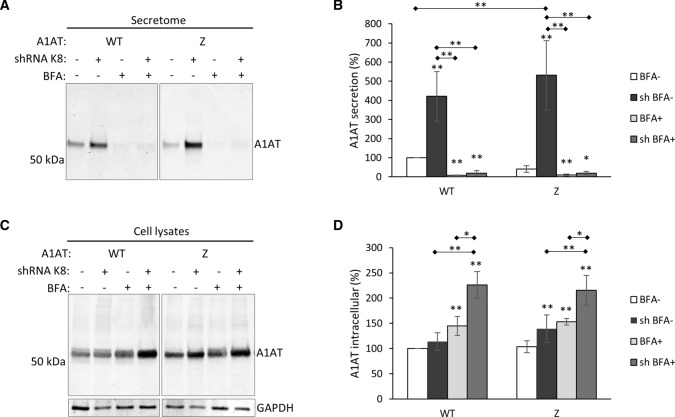
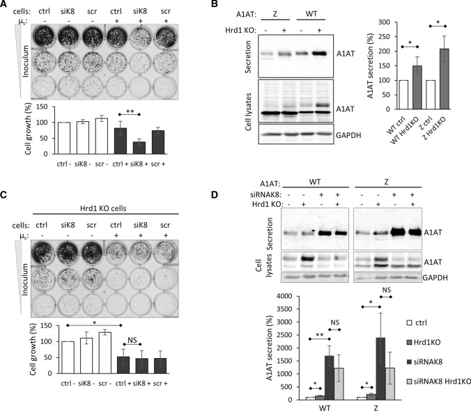
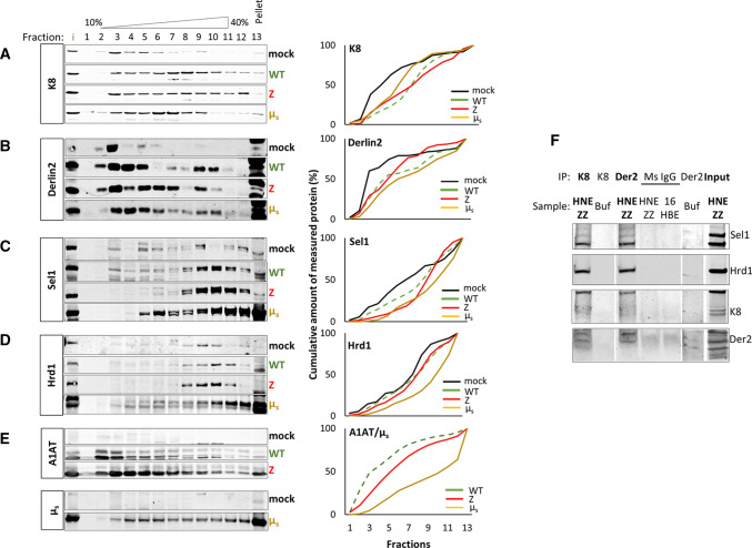
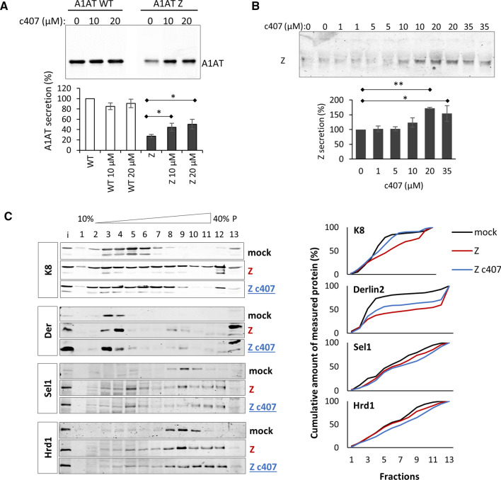
Early recognition and enhanced degradation of misfolded proteins by the endoplasmic reticulum (ER) quality control and ER-associated degradation (ERAD) cause defective protein secretion and membrane targeting, as exemplified for Z-alpha-1-antitrypsin (Z-A1AT), responsible for alpha-1-antitrypsin deficiency (A1ATD) and F508del-CFTR (cystic fibrosis transmembrane conductance regulator) responsible for cystic fibrosis (CF). Prompted by our previous observation that decreasing Keratin 8 (K8) expression increased trafficking of F508del-CFTR to the plasma membrane, we investigated whether K8 impacts trafficking of soluble misfolded Z-A1AT protein. The subsequent goal of this study was to elucidate the mechanism underlying the K8-dependent regulation of protein trafficking, focusing on the ERAD pathway. The results show that diminishing K8 concentration in HeLa cells enhances secretion of both Z-A1AT and wild-type (WT) A1AT with a 13-fold and fourfold increase, respectively. K8 down-regulation triggers ER failure and cellular apoptosis when ER stress is jointly elicited by conditional expression of the µs heavy chains, as previously shown for Hrd1 knock-out. Simultaneous K8 silencing and Hrd1 knock-out did not show any synergistic effect, consistent with K8 acting in the Hrd1-governed ERAD step. Fractionation and co-immunoprecipitation experiments reveal that K8 is recruited to ERAD complexes containing Derlin2, Sel1 and Hrd1 proteins upon expression of Z/WT-A1AT and F508del-CFTR. Treatment of the cells with c407, a small molecule inhibiting K8 interaction, decreases K8 and Derlin2 recruitment to high-order ERAD complexes. This was associated with increased Z-A1AT secretion in both HeLa and Z-homozygous A1ATD patients' respiratory cells. Overall, we provide evidence that K8 acts as an ERAD modulator. It may play a scaffolding protein role for early-stage ERAD complexes, regulating Hrd1-governed retrotranslocation initiation/ubiquitination processes. Targeting K8-containing ERAD complexes is an attractive strategy for the pharmacotherapy of A1ATD.

Keywords: Cytoskeleton; Epithelium; Intermediary filaments; Protein complexes fractionation; Protein–protein interaction; Synthetic lethality.


Conflict of interest statement

The authors have no relevant financial or non-financial interests to disclose.

Figures

Fig. 1
Fig. 1
Silencing K8 in HeLa cells increases the secretion of Z-A1AT and WT-A1AT through conventional secretory pathway. A WB analysis of secretion of WT-A1AT/Z-A1AT in cell cultures with normal (shRNAK8-) and decreased (shRNAK8 +) level of K8 expression after treatment with Brefeldin A 5 µg/ml (BFA +) or vehicle (BFA−) for 7 h. Representative image and quantification from n = 9 independent experiments. B Quantification of secretion (mean ± SD) normalized to total protein concentrations in respective cell lysates from at least four experiments. *< 0.05; **< 0.005; ***< 0.0005 (Mann–Whitney test); *over the bar indicates p value vs. control (white boxes), if otherwise it is indicated. C WB analysis of intracellular WT-A1AT/Z-A1AT in cell cultures with normal (shRNAK8-) and decreased (shRNAK8 +) level of K8 expression after treatment with Brefeldin A 5 µg/ml (BFA +) or vehicle (BFA−) for 7 h. Representative image and quantification from n = 9 independent experiments. D Quantification of secretion (mean ± SD) normalized to total protein concentrations in respective cell lysates from at least 4 experiments. *< 0.05; **< 0.005; ***< 0.0005 (Mann–Whitney test); * over the bar indicates p value vs. control (white boxes), if otherwise it is indicated
Fig. 2
Fig. 2
K8 regulates Hrd1-dependent processes. A Synthetic lethality of HeLa cells conditionally expressing µs. Cells transfected with siRNA against K8 (cells siK8), or scrambled siRNA (cells scr) were treated with 0.5 nM Mifepristone (Mif) (µs +) or vehicle (µs−). After transfection cells were seeded upon 1:5 serial dilution (inoculum 5000, 1000, 200 cells) into 24-well plates and grown for 7 days. Scrambled RNA had no effect on cell growth of non-expressing (scr-µs) and µs-expressing cells (scr + µs). This is a representative image of a culture plate and quantification of cell growth (inoculum 1000 cells, middle row) as mean % (± SD) of growth in non-transfected non-treated cells (ctrl-) of five independent experiments are shown. **p < 0.005 (paired t test). B Effect of Hrd1 knock-out on WT-A1AT and Z-A1AT secretion from HeLa cells. WB images show WT-A1AT and Z-A1AT secretion levels (upper panel), A1AT expression (middle panel) and GAPDH as loading control (lower panel). Protein quantification is expressed as means ± SD from four independent experiments. *p < 0.05 (Mann–Whitney test). C Synthetic lethality of Hrd1KO HeLa cells. Cells were treated as in (A). Representative image of culture plate and quantification of cell growth (inoculum 1000 cells, middle row) as mean % (± SD) of growth in non-transfected non-treated cells (ctrl-) of five independent experiments are shown. *p < 0.05 (paired t test). D Effects of K8 silencing, Hrd1 knock-out and their combination on secretion of WT-A1AT and Z-A1AT. Normal (Hrd1KO−) and Hrd1KO (Hrd1KO +) HeLa cells were transiently transfected with WT-A1AT or Z-A1AT and with siRNA against K8 (siRNAK8 +). Representative WB images demonstrating WT-A1AT and Z-A1AT secretion (upper image), WT-A1AT and Z-A1AT expression (middle image) and GAPDH (bottom image) from normal, Hrd1KO + , siRNAK8 + and siRNAK8 + /Hrd1KO + HeLa cells. Protein quantification of A1AT secretion is expressed as mean ± SD for three experiments. *p < 0.05, **p < 0.005 (Mann–Whitney test)
Fig. 3
Fig. 3
K8 is recruited to the ERAD complexes upon overexpression of misfolded Z-A1AT, WT-A1AT and µs. Sucrose gradient fractionation of the ERAD complexes. HeLa cells expressing WT-A1AT (WT-A1AT), Z-A1AT (Z-A1AT), µs (Mif 0.5 nM for 24 h) (µs), or mock-transfected (mock). Protein samples from cells were sedimented over a 10–40% sucrose gradient. K8 (A), Sel1 (B), Hrd1 (C), Derlin-2 (D), and µs/WT-A1AT/Z-A1AT (E) were detected by immunoblotting with respective antibodies and semi-quantified (right panels). Quantifications represent results shown in the corresponding images on the left and are expressed as cumulative amount of measured protein (%) to demonstrate the overall shift. In the right panel: black lines correspond to protein content in low density ERAD complexes under basal conditions, green, red, and yellow lines show ERAD complexes formed upon WT-A1AT, Z-A1AT and µs expression, respectively. The three tested substrates distributed differently in sucrose gradients: a large amount of µs which is a substrate efficiently processed by ERAD strongly distributed to heavy fractions, a relatively large amount of Z-A1AT distributed to heavy fractions, whereas smaller amounts of WT-A1AT were recruited to heavy fractions. Experiments were performed at least three times for all tested conditions. F Co-immunoprecipitation of ERAD complexes from microsomes of polarized primary human nasal epithelial cells homozygous for Z mutation of A1AT. Derlin2 or K8 were immunoprecipitated with anti-Derlin2 or anti-K8 antibodies, respectively (see Methods section) and proteins of interest were detected using corresponding antibodies (Sel1, Hrd1, K8, Derlin2). Negative controls with unspecific mouse IgG (Ms IgG) and extracts, as well as with anti-Derlin2/K8 antibody and LMNG buffer were applied
Fig. 4
Fig. 4
Silencing K8 affects ERAD complexes formation. Sucrose gradient fractionation of the ERAD complexes. HeLa cells expressing normal (mock and Z-A1AT) and decreased level of K8 (shK8 and shK8 Z-A1AT). Protein samples from cells were sedimented over a 10–40% sucrose gradient. K8 (A), Sel1 (B), Hrd1 (C), Derlin-2 (D) were detected by immunoblotting with respective antibodies. Black arrows correspond to ERAD complexes containing K8 and Derlin2 formed in shK8 cells, red dashed lined boxes correspond to high-density ERAD complexes formed in presence of K8 and Z-A1AT (Z-A1AT cells) but missing in shK8 Z-A1AT cells. Quantifications represent results shown in the corresponding images on the left and are expressed as cumulative amount of measured protein (%) to demonstrate the overall shift
Fig. 5
Fig. 5
K8 is recruited to the ERAD complexes upon overexpression of misfolded F508del-CFTR. 16HBE cells expressing WT-CFTR and F508del-CFTR, as indicated, were lysed in 1% w/v LMNG and sedimented over a 10–40% w/v sucrose gradient. Levels of K8 (Aa and Ab), Hrd1 (D), Sel1 (C), Derlin-2 (B), were detected by immunoblotting. Representative images are shown. Quantifications represent results shown in the corresponding images on the left and are expressed as cumulative amount of measured protein (%) to demonstrate the overall shift. Experiment was performed three times. E Co-immunoprecipitation of ERAD complexes from microsomes of 16HBE cells expressing WT- or F508del-CFTR. Derlin2 or K8 were immunoprecipitated with anti-Derlin2 (top panel) or anti-K8 antibodies (bottom panel), respectively (see Methods section) and proteins of interest were detected using corresponding antibodies (Sel1, Hrd1, K8, Derlin2). Negative controls with unspecific mouse IgG (Ms IgG) and extracts, as well as with anti-Derlin2/K8 antibody and LMNG buffer were applied
Fig. 6
Fig. 6
Compound c407 increases secretion of Z-A1AT from HeLa and primary HNE cells through modulating K8-contianing ERAD complexes. A Quantification of WT-A1AT and Z-A1AT secretion level from HeLa cells upon treatment with c407 (10 and 20) and vehicle (0). Cells were treated with c407 at concentrations of 10 and 20 µM for 24 h. The representative WB analysis image shows A1AT secreted into the medium. B Quantification of Z-A1AT secretion level from primary HNE cells upon treatment with c407 (1, 5, 10, 20 and 35) and vehicle (0). Cells were treated with c407 at concentrations of 1, 5, 10, 20 and 35 µM f or 14 days as pre-treatment and for 24 h for test of secretion. The representative WB analysis image shows A1AT secreted into the medium. C Sucrose gradient fractionation results upon c407 treatment. HeLa cells were transfected with Z-A1AT (Z) and treated with c407 at concentration of 20 µM or vehicle for 48 h, as indicated. Levels of K8, Hrd1, Sel1, and Derlin-2 were detected by immunoblotting in all fractions. Representative images are shown. Quantifications represent results shown in the corresponding images on the left and are expressed as cumulative amount of measured protein (%) to demonstrate the overall shift. Experiment was performed three times
Fig. 7
Fig. 7
Model of K8 recruitment to the ERAD complexes at the ER membrane—hypothesis (Adapted from Christianson and Ye). Normal K8 recruitment to the ERAD complexes: A K8 as a scaffolding platform in constant complex with Derlin2. Z-A1AT is recognized and recruited for retrotranslocation and ubiquitination. Stage rescuable for Z-A1AT secretion (upon Hrd1 or K8 down-regulation). Low density fractions complexes containing Derlin2 (module 2A in Supp. Fig. 1). B K8 facilitates complexing with Hrd1/Sel1. Retrotranslocation of ubiquitinated Z-A1AT is initiated. Stage rescuable for Z-A1AT secretion (upon Hrd1 or K8 down-regulation). Medium to high-density fractions complexes containing Derlin2, Sel1 and Hrd1 (modules 2A + 2B in Supp. Fig. 1). C Z-A1AT is being dislocated from the ER lumen to the cytosol. Dislocation stage no rescuable for Z-A1AT secretion (upon p97 inhibition). Hypothetic higher-order complexes containing Derlin2, Sel1, Hrd1 and p97. Decreased or no recruitment of K8 to the ERAD complexes upon c407 molecule treatment or siRNA against K8: D K8–Derlin2 complex maintained (low-density fractions) even with residual K8. However, K8-dependent scaffolding of higher-order complexes is affected. Recruitment of Z-A1AT for retrotranslocation and ubiquitination is reduced. ER-luminal Z-A1AT is secreted. E K8–Derlin2 is not recruited to form complexes with Hrd1/Sel1. The shRNAK8 decreases Sel1 and Hrd1 recruitment to high-order complexes. By contrast, c407 treatment of cells do not change Hrd1 and Sel1 recruitment to higher-order complexes. Retrotranslocation initiation and ubiquitination of Z-A1AT is reduced. ER-luminal Z-A1AT is secreted. F Z-A1AT dislocation to the cytosol is reduced. ER-luminal Z-A1AT is secreted

References

    1. Bakunts A, Orsi A, Vitale M et al (2017) Ratiometric sensing of BiP-client versus BiP levels by the unfolded protein response determines its signaling amplitude. Elife 6:e27518. 10.7554/eLife.27518 - PMC - PubMed
    1. Baldridge RD, Rapoport TA (2016) Autoubiquitination of the Hrd1 ligase triggers protein retrotranslocation in ERAD. Cell 166:394–407. 10.1016/j.cell.2016.05.048 - PMC - PubMed
    1. Besingi RN, Clark PL (2015) Extracellular protease digestion to evaluate membrane protein cell surface localization. Nat Protoc 10:2074–2080. 10.1038/nprot.2015.131 - PMC - PubMed
    1. Carlson EJ, Pitonzo D, Skach WR (2006) p97 functions as an auxiliary factor to facilitate TM domain extraction during CFTR ER-associated degradation. EMBO J 25:4557–4566. 10.1038/sj.emboj.7601307 - PMC - PubMed
    1. Chakraborty P, Teckman J (2014) Alpha-1-antitrypsin deficiency liver disease: science and therapeutic potential 50 years later. J Gastroenterol Pancreatol Liver Disord 1(3):1–9. 10.15226/2374-815X/1/3/00113

MeSH terms

Substances

LinkOut - more resources