. 2022 Aug 31;19(1):210.
doi: 10.1186/s12974-022-02575-y.

Chronic allergic lung inflammation negatively influences neurobehavioral outcomes in mice

Affiliations

Chronic allergic lung inflammation negatively influences neurobehavioral outcomes in mice

Akihiro Kanaya et al. J Neuroinflammation. .

Abstract

Background: Asthma is a major public health problem worldwide. Emerging data from epidemiological studies show that allergies and allergic diseases may be linked to anxiety, depression and cognitive decline. However, little is known about the effect of asthma, an allergic lung inflammation, on cognitive decline/behavioral changes. Therefore, we investigated the hypothesis that allergic lung inflammation causes inflammation in the brain and leads to neurobehavioral changes in mice.

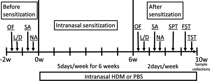
Methods: Wild-type C57BL/6J female mice were sensitized with nasal house dust mite (HDM) antigen or control PBS for 6 weeks to induce chronic allergic lung inflammation. A series of neurocognitive tests for anxiety and/or depression were performed before and after the intranasal HDM administration. After the behavior tests, tissues were harvested to measure inflammation in the lungs and the brains.

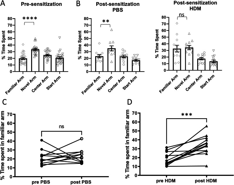
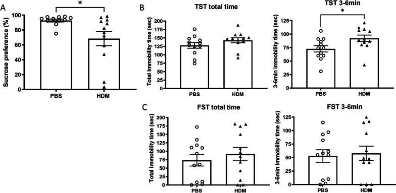
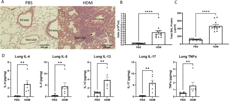
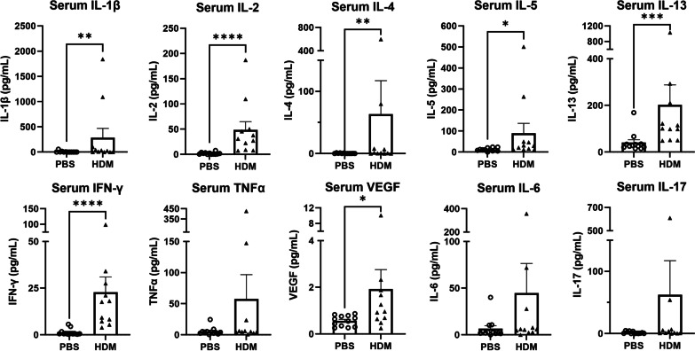
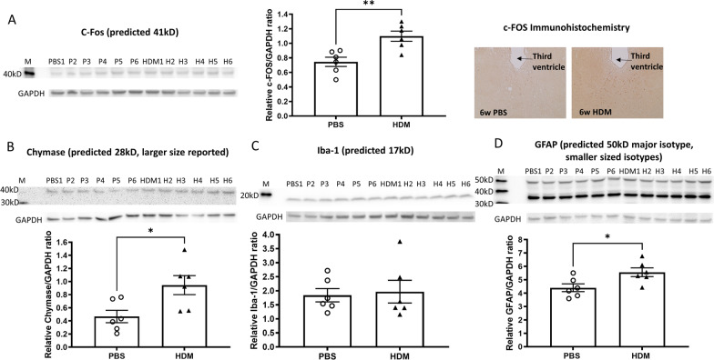
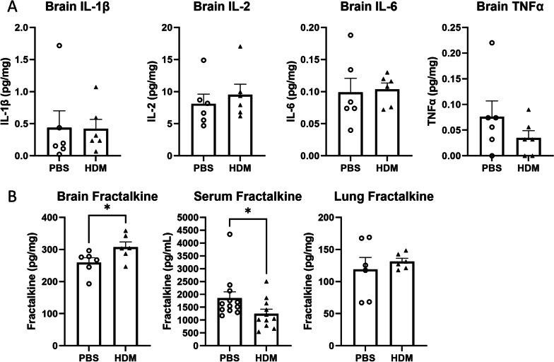
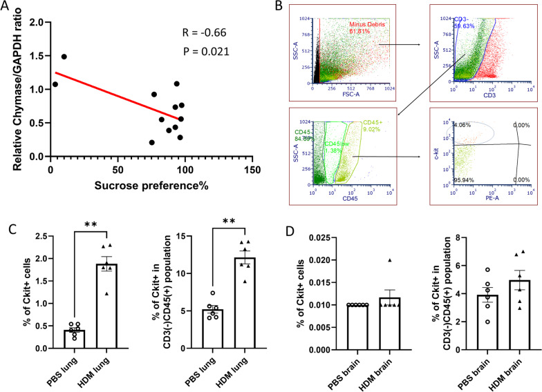
Results: HDM-treated mice exhibited significantly increased immobility times during tail suspension tests and significantly decreased sucrose preference compared with PBS controls, suggesting a more depressed and anhedonia phenotype. Spatial memory impairment was also observed in HDM-treated mice when assessed by the Y-maze novel arm tests. Development of lung inflammation after 6 weeks of HDM administration was confirmed by histology, bronchoalveolar lavage (BAL) cell count and lung cytokine measurements. Serum pro-inflammatory cytokines and Th2-related cytokines levels were elevated in HDM-sensitized mice. In the brain, the chemokine fractalkine was increased in the HDM group. The c-Fos protein, a marker for neuronal activity, Glial Fibrillary Acidic Protein (GFAP) and chymase, a serine protease from mast cells, were increased in the brains from mice in HDM group. Chymase expression in the brain was negatively correlated with the results of sucrose preference rate in individual mice.

Conclusions: 6 weeks of intranasal HDM administration in mice to mimic the chronic status of lung inflammation in asthma, caused significant inflammatory histological changes in the lungs, and several behavioral changes consistent with depression and altered spatial memory. Chymase and c-Fos proteins were increased in the brain from HDM-treated mice, suggesting links between lung inflammation and brain mast cell activation, which could be responsible for depression-like behavior.

Keywords: Asthma; Chronic allergic lung inflammation; Depression; Mast cell; Neurobehavior; Spatial memory.


Conflict of interest statement

The authors declare they have no competing interests.

Figures

Fig. 1
Fig. 1
Schematic representation of the timeline of experiments. Before sensitization, OF, L/D, SA and NA were performed. Mice were sensitized by intranasal administration of house dust mite (HDM) or control PBS, 5 days/wk for 6 weeks. After sensitization, mice underwent behavioral testing using the OF, L/D, SA, NA, FST, TST and Sucrose preference test. The interval between each behavioral test was 2–3 days. 1. OF; open field, 2. L/D; light/dark transition test, 3. SA; Y-maze spontaneous alternation test, 4. NA; Y-maze novel arm test, 5. FST; forced swim test, 6. TST; tail suspension test. 7. SPT; sucrose preference test
Fig. 2
Fig. 2
Locomotor activity measured by the open field test before and after 6 weeks of intranasal HDM or PBS (control). A, B The ambulatory distance over each 10-min time bin during the 60-min trial before (n = 24, total mice) and after (n = 12 per group) intranasal sensitization. *p < 0.05, comparing exploratory activity over the duration of the session, between treatment groups, two-way ANOVA. C, D A comparison of the total ambulatory distance in individual mice, pre- and post- intranasal sensitization with HDM or PBS (control). Mice in the HDM group did not show a typical reduction of ambulatory distances traveled after sensitization. E, F A comparison of the total time spent in the center of the open field chamber in individual mice (n = 12 each, *p < 0.05, ***p < 0.001, Wilcoxon paired t-test)
Fig. 3
Fig. 3
Exploratory behavior and the animal’s preference for the light measured in the light–dark transition test. A Before initiation of nasal PBS or HDM treatment, mice spent more time in the dark side. B After PBS treatment, control mice maintained a preference for the dark while HDM-sensitized mice spent about the same time in the light and dark chambers, which is unusual considering rodents’ natural aversion to bright spaces. ****p < 0.0001, Mann–Whitney. C, D The time ratio spent in the dark versus the light in individual mice before and after the sensitization was significantly lower in HDM group but not in the control PBS group. **p < 0.01, Wilcoxon paired t-test. E, F The number of transitions (how often the light–dark transition zone was crossed) in individual mice was remarkably higher in the HDM group after sensitization. ***p < 0.001, Wilcoxon paired t-test
Fig. 4
Fig. 4
Spatial memory assessed by the Y-maze novel arm test. A Before sensitization, mice spent more time in the novel arm compared to the familiar arm. B After treatment, control mice again spent more time in the novel arm than the familiar arm, however, the HDM-sensitized mice spent about the same time in the familiar arm, indicating poor spatial memory. ****p < 0.0001, **p < 0.01, ns not significant, one-way ANOVA novel vs. familiar. C, D A comparison of the percent time spent in the familiar arm in individual mice. Only mice in the HDM group showed an increased time spent in the familiar arm, again indicating impaired spatial memory. ***p < 0.001, Wilcoxon paired t-test
Fig. 5
Fig. 5
Depression-like behaviors assessed by the sucrose preference test, the tail suspension test and the forced swim test. A Mice treated with intranasal HDM exhibited decreased sucrose preference compared with PBS control mice. *p < 0.05, Mann–Whitney. B After 6 weeks of intranasal PBS (control) or HDM administration, mice underwent the tail suspension test (TST) and the duration of no movement (immobility time) was recorded. The HDM group showed increased immobility time during the last 3 min, indicating increased depressive-like behavior (*p < 0.05, Mann–Whitney). C In the forced swim test (FST), no significant difference was observed between mice in the PBS control versus HDM sensitization group
Fig. 6
Fig. 6
Six weeks of nasal HDM administration caused lung inflammation measured by histology, BAL cell counts, BAL total protein concentrations and lung cytokine measurements. A Six weeks of intranasal HDM administration caused marked peri-bronchial and peri-vascular infiltration of mononuclear cells. B Total cells counted in the bronchoalveolar lavage (BAL) were significantly higher in the HDM group. C Protein concentrations in the BAL were elevated in the HDM group. D Multiple pro-inflammatory cytokine concentrations in lung digests were higher in the HDM group compared to PBS control. **p < 0.01, ****p < 0.0001, Mann–Whitney
Fig. 7
Fig. 7
Cytokines measured in the serum after 6 weeks of nasal PBS or HDM sensitization. After chronic intranasal HDM sensitization, changes in serum cytokine concentrations were observed. *p < 0.05, **p < 0.01, ***p < 0.001, ****p < 0.0001, Mann–Whitney
Fig. 8
Fig. 8
Brain pro-inflammatory and cell activation-associated protein levels. A C-Fos levels were elevated in the brain from HDM-sensitized mice, assessed by immunoblotting and immunohistochemistry of paraffin section (brown dots, prominent in the periventricular thalamus). BD Chymase, Iba-1 and GFAP expression measured by immunoblotting of brain homogenates. GAPDH was used to normalize potential variation in protein loading on gels. Chymase and GFAP protein were increased in the whole brains of HDM-treated mice. *p < 0.05, **p < 0.01, Mann–Whitney
Fig. 9
Fig. 9
Concentrations of cytokines and chemokines in the whole brain of mice after intranasal sensitization. A After completion of behavioral tests, inflammatory cytokines in the whole brain were measured. B Fractalkine concentrations in the whole brain, serum and lungs. *p < 0.05, Mann–Whitney
Fig. 10
Fig. 10
A Correlation between chymase expression in the brain and sucrose preference in individual mice. B A representative gating used to identify mast cells. C, D C-kit positive mast cells in the lungs and brain, expressed as % of total singlet cells (left) and in CD3 negative, CD45 positive population (right). **p < 0.01, Mann–Whitney

References

    1. Backman H, Raisanen P, Hedman L, Stridsman C, Andersson M, Lindberg A, et al. Increased prevalence of allergic asthma from 1996 to 2006 and further to 2016-results from three population surveys. Clin Exp Allergy. 2017;47(11):1426–1435. doi: 10.1111/cea.12963. - DOI - PubMed
    1. Moore WC, Bleecker ER, Curran-Everett D, Erzurum SC, Ameredes BT, Bacharier L, et al. Characterization of the severe asthma phenotype by the National Heart, Lung, and Blood Institute's Severe Asthma Research Program. J Allergy Clin Immunol. 2007;119(2):405–413. doi: 10.1016/j.jaci.2006.11.639. - DOI - PMC - PubMed
    1. Richardson LP, Lozano P, Russo J, McCauley E, Bush T, Katon W. Asthma symptom burden: relationship to asthma severity and anxiety and depression symptoms. Pediatrics. 2006;118(3):1042–1051. doi: 10.1542/peds.2006-0249. - DOI - PubMed
    1. Opolski M, Wilson I. Asthma and depression: a pragmatic review of the literature and recommendations for future research. Clin Pract Epidemiol Ment Health. 2005;1:18. doi: 10.1186/1745-0179-1-18. - DOI - PMC - PubMed
    1. Walters P, Schofield P, Howard L, Ashworth M, Tylee A. The relationship between asthma and depression in primary care patients: a historical cohort and nested case control study. PLoS ONE. 2011;6(6):e20750. doi: 10.1371/journal.pone.0020750. - DOI - PMC - PubMed

MeSH terms