. 2022 Aug 31;10(1):141.
doi: 10.1186/s40168-022-01337-5.

Lactic acid from vaginal microbiota enhances cervicovaginal epithelial barrier integrity by promoting tight junction protein expression

Affiliations

Lactic acid from vaginal microbiota enhances cervicovaginal epithelial barrier integrity by promoting tight junction protein expression

David Jose Delgado-Diaz et al. Microbiome. .

Abstract

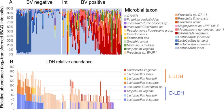
Background: Women with a cervicovaginal microbiota dominated by Lactobacillus spp. are at reduced risk of acquiring sexually transmitted infections including HIV, but the biological mechanisms involved remain poorly defined. Here, we performed metaproteomics on vaginal swab samples from young South African women (n = 113) and transcriptomics analysis of cervicovaginal epithelial cell cultures to examine the ability of lactic acid, a metabolite produced by cervicovaginal lactobacilli, to modulate genital epithelial barrier function.

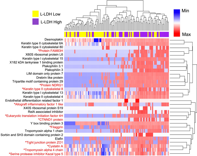
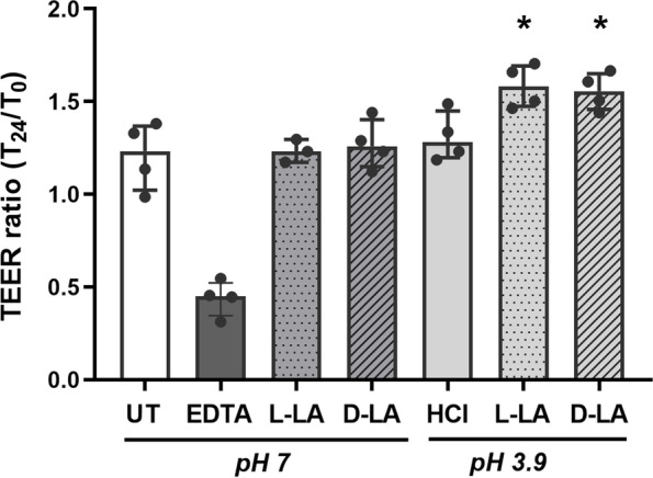
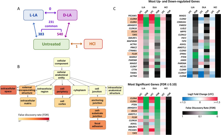
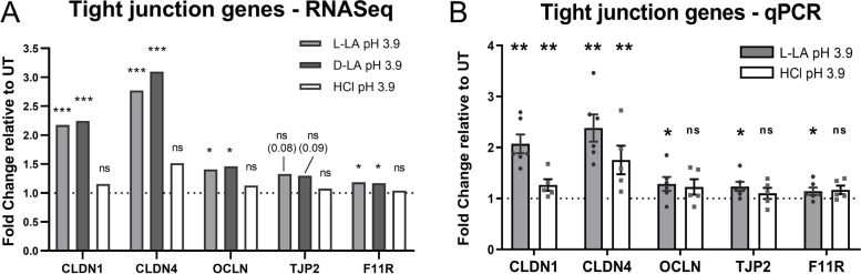
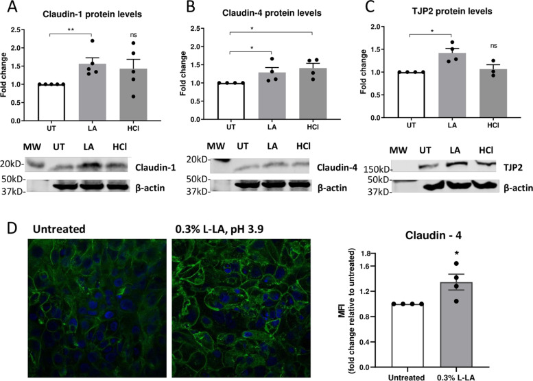
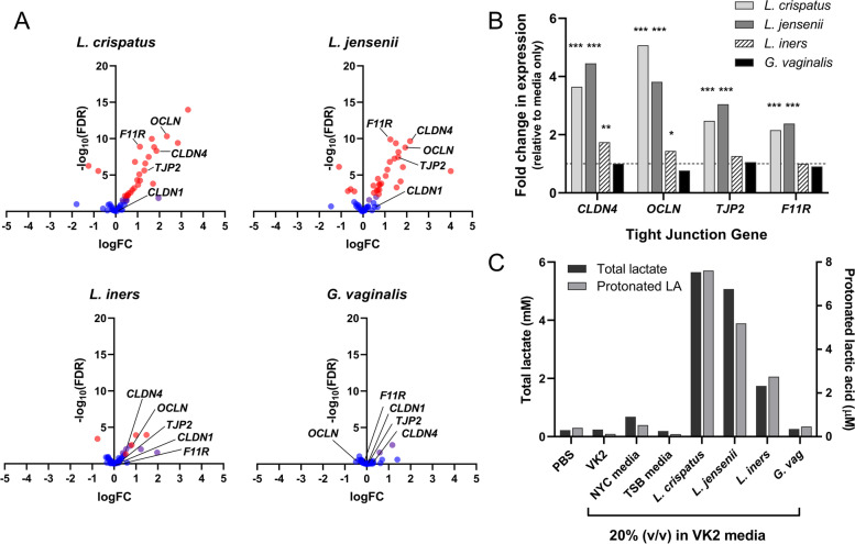
Results: Compared to women with Lactobacillus-depleted microbiota, women dominated by vaginal lactobacilli exhibit higher abundance of bacterial lactate dehydrogenase, a key enzyme responsible for lactic acid production, which is independently associated with an increased abundance of epithelial barrier proteins. Physiological concentrations of lactic acid enhance epithelial cell culture barrier integrity and increase intercellular junctional molecule expression.

Conclusions: These findings reveal a novel ability of vaginal lactic acid to enhance genital epithelial barrier integrity that may help prevent invasion by sexually transmitted pathogens. Video abstract.

Keywords: Epithelial cells; Female reproductive tract; HIV; Lactic acid; Lactobacilli; Metabolites; STIs; Tight junctions; Transcriptomics; Vaginal microbiome.


Conflict of interest statement

GT and ACH are coinventors on a patent application examining the immunomodulatory effects of L-lactic acid on cervicovaginal epithelial cells. The other authors declare that they have no competing interests.

Figures

Fig. 1
Fig. 1
Higher relative abundance of bacterial LDH in women with a Lactobacillus-dominated microbiome. The vaginal microbiome composition of 113 young women from Cape Town, South Africa, was determined using liquid chromatography-tandem mass spectrometry, and participants were classified as being bacterial vaginosis (BV) negative, positive, or intermediate (Int) using Nugent-BV criteria. Proteins were identified using MaxQuant, and a custom database was generated using de novo sequencing to filter the UniProt database. Taxonomy was assigned using UniProt, and relative abundance of each taxon was determined by aggregating the intensity-based absolute quantification (iBAQ) values of all proteins identified for each taxon. The relative abundance of the 20 most abundant bacterial species is indicated for each participant in (A). The relative abundance of L- and D-lactate dehydrogenase (LDH) protein derived from various bacterial sources was measured in vaginal secretions using metaproteomic analyses for each participant (B)
Fig. 2
Fig. 2
L-LDH relative abundance in the FRT is associated with expression of epithelial junction proteins. Unsupervised hierarchical clustering of intensity-based absolute quantification (iBAQ) values for epithelial barrier-related proteins that were found to be differentially abundant between women (n = 113) with levels of L-LDH protein above (high, purple) or below (low, yellow) the median value for the cohort. The moderated t-test (limma package, R) was used to identify proteins significantly associated with L-LDH relative abundance following false discovery rate adjustment for multiple comparisons. *Proteins that remained significantly associated with L-LDH after adjusting for Lactobacillus relative abundance, as well as the presence of sexually transmitted infections, contraceptive use, and detection of prostate-specific antigen
Fig. 3
Fig. 3
Protonated LA increases the barrier integrity of ectocervical epithelial cells. Ectocervical cells (Ect1) seeded into transwells were apically treated for 1 h with media alone (untreated, UT) or media containing 0.3% L- or D-lactic acid (LA) at either low (pH 3.9) or neutral (pH 7) pH, low pH media alone (HCl, pH 3.9), or 2 mM EDTA. Epithelial barrier integrity was assessed by transepithelial electrical resistance (TEER) measured at baseline (T0) and 24 h after treatment (T24) expressed as the TEER ratio (T24/T0). Graph shows median and interquartile range for each condition from 4 independent experiments. *p < 0.05 compared to untreated control, as determined by Mann-Whitney U-test
Fig. 4
Fig. 4
LA treatment of ectocervical epithelial cells increases the expression of genes relevant to barrier integrity. Ectocervical epithelial cells were apically treated with 0.3% L- or D-lactic acid (LA) or low pH media alone (HCl) for 1 h and transcriptomic changes assessed at 4-h post treatment by RNA-Seq. A Number of genes differentially expressed between the treatments is shown next to arrows indicating comparison groups as assessed by voom-limma analysis (Log2 fold change > 0.5, false discovery rate [FDR] < 0.05). There were 231 genes which were differentially expressed by both L-LA and D-LA as compared to untreated cells. B Gene ontologies enriched by LA treatment. For visual simplicity, ontologies enriched with a FDR < 0.01 are shown; all ontologies enriched with a FDR < 0.05 are shown in Supplementary Table 2. C Expression of genes related to epithelial tight junctions relative to untreated cells ranked by magnitude of up- or downregulation (left panel) or adjusted p-value (FDR). Genes highlighted in orange were those selected for further characterisation
Fig. 5
Fig. 5
qRT-PCR analysis of expression of tight junction genes in ectocervical cells following L-LA treatment. Ectocervical epithelial cells were apically treated with 0.3% L-lactic acid (L-LA) or low pH media alone (HCl) for 1 h, and gene expression of tight junction factors claudin-1 (CLDN1), claudin-4 (CLDN4), occludin (OCLN), zona occludens-2/tight junction protein-2 (TJP2) and F11R was assessed at 4 h post treatment by RNA-Seq (A) or qRT-PCR (B). A Fold change in expression of tight junction genes in ectocervical epithelial cells treated with L-LA, D-LA or HCl-treated cells relative to untreated, as determined by RNA-Seq analysis. * and *** represent false discovery rate < 0.05 and < 0.001, respectively. B Relative expression (fold change) of tight junction genes compared to untreated cells (indicated by dotted line) as determined by qRT-PCR. Graph shows mean +/− SEM from n = 5–6 independent experiments. * and ** represent p < 0.05 and < 0.01, respectively, compared to untreated cells as determined by the Mann-Whitney U-test. NS, not significant
Fig. 6
Fig. 6
Increased expression of tight junction proteins in ectocervical cells by L-LA. Protein levels of barrier proteins claudin-1 (A), claudin-4 (B) and tight junction protein-2 (TJP2) (C) in ectocervical epithelial cells stimulated apically with 0.3% L-LA pH 3.9 (LA) or low pH media (HCl, pH 3.9) for 1 h followed by lysis of cells at 24 h and assessed by Western blot analysis. Target protein levels were standardised to β-actin in the same sample. D Expression of claudin-4 was visualised in ectocervical epithelial cells treated as above by immunofluorescence and confocal microscopy. Images from a single experiment representative of n = 4 replicates are shown (left), and mean fluorescence intensity (MFI) of claudin-4 signal intensity was assessed using ImageJ software and expressed relative to untreated samples. All graphs show mean +/− SEM (expressed as fold change compared to untreated cells) from 3 to 5 independent experiments. Significance was assessed by the Mann-Whitney U-test; *, **p < 0.05 and 0.01, respectively
Fig. 7
Fig. 7
Lactobacillus cultures producing high levels of LA upregulate tight junction gene expression. VK2 epithelial cells were treated with media containing 20% (v/v) filtered supernatant from cultures of L. crispatus, L. jensenii, L. iners or G. vaginalis bacteria. The impact of treatment on expression of key tight junction genes was analysed by RNA-Seq, and volcano plots of changes in expression following treatment are shown in (A), with red dots indicating significantly DEGs (FDR < 0.05). B Fold change in expression of claudin-4 (CLDN4), occludin (OCLN), zona occludens-2/tight junction protein-2 (TJP2) and F11R genes relative to cells treated with media alone (mean, from n = 3 replicates; *, ** and ***false discovery rate < 0.5, 0.01 and 0.001, respectively). C Concentration of total lactate (black bars) and protonated LA (grey bars) in treatment media, the latter calculated using the Henderson-Hasselbalch equation and the treatment media pH (range 6.6–7.1). Values were normalised to account for slight variations in growth rate of bacterial cultures

Similar articles

Cited by

References

    1. Tuddenham S, Ravel J, Marrazzo JM. Protection and risk: Male and female genital microbiota and sexually transmitted infections. J Infect Dis. 2021;223(Supplement_3):S222–SS35. doi: 10.1093/infdis/jiaa762. - DOI - PMC - PubMed
    1. Ravel J, Gajer P, Abdo Z, Schneider GM, Koenig SS, McCulle SL, et al. Vaginal microbiome of reproductive-age women. Proc Natl Acad Sci U S A. 2011;108(Suppl 1):4680–4687. doi: 10.1073/pnas.1002611107. - DOI - PMC - PubMed
    1. Fettweis JM, Brooks JP, Serrano MG, Sheth NU, Girerd PH, Edwards DJ, The Vaginal Microbiome Consortium. Jefferson KK, Buck GA, et al. Differences in vaginal microbiome in African American women versus women of European ancestry. Microbiology (Reading) 2014;160(Pt 10):2272–2282. doi: 10.1099/mic.0.081034-0. - DOI - PMC - PubMed
    1. Aldunate M, Srbinovski D, Hearps AC, Latham CF, Ramsland PA, Gugasyan R, et al. Antimicrobial and immune modulatory effects of lactic acid and short chain fatty acids produced by vaginal microbiota associated with eubiosis and bacterial vaginosis. Front Physiol. 2015;6:164. doi: 10.3389/fphys.2015.00164. - DOI - PMC - PubMed
    1. McKinnon LR, Achilles SL, Bradshaw CS, Burgener A, Crucitti T, Fredricks DN, et al. The evolving facets of bacterial vaginosis: implications for HIV transmission. AIDS Res Hum Retroviruses. 2019;35:219–228. doi: 10.1089/aid.2018.0304. - DOI - PMC - PubMed

Publication types

LinkOut - more resources