. 2022 Aug 31;15(1):123.
doi: 10.1186/s13045-022-01343-y.

Comparative analysis of cancer cell responses to targeted radionuclide therapy (TRT) and external beam radiotherapy (EBRT)

Affiliations

Comparative analysis of cancer cell responses to targeted radionuclide therapy (TRT) and external beam radiotherapy (EBRT)

Michal Grzmil et al. J Hematol Oncol. .

Abstract

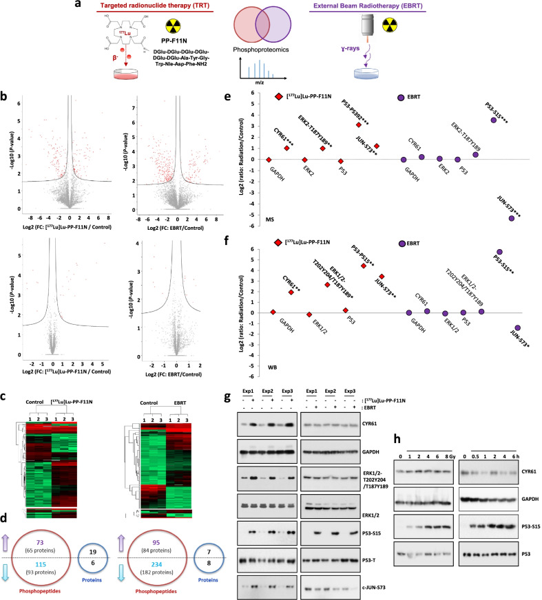
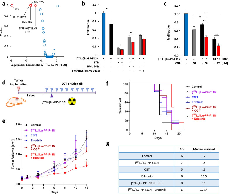
The vast majority of our knowledge regarding cancer radiobiology and the activation of radioresistance mechanisms emerged from studies using external beam radiation therapy (EBRT). Yet, less is known about the cancer response to internal targeted radionuclide therapy (TRT). Our comparative phosphoproteomics analyzed cellular responses to TRT with lutetium-177-labeled minigastrin analogue [177Lu]Lu-PP-F11N (β-emitter) and EBRT (ɣ-rays) in CCKBR-positive cancer cells. Activation of DNA damage response by p53 was induced by both types of radiotherapy, whereas TRT robustly increased activation of signaling pathways including epidermal growth factor receptor (EGFR), mitogen-activated protein kinases (MAPKs) or integrin receptor. Inhibition of EGFR or integrin signaling sensitized cancer cells to radiolabeled minigastrin. In vivo, EGFR inhibitor erlotinib increased therapeutic response to [177Lu]Lu-PP-F11N and median survival of A431/CCKBR-tumor bearing nude mice. In summary, our study explores a complex scenario of cancer responses to different types of irradiation and pinpoints the radiosensitizing strategy, based on the targeting survival pathways, which are activated by TRT.

Keywords: CCKBR; Erlotinib; Minigastrin; Phosphoproteomics; Radioresistance.


Conflict of interest statement

MB and RS are inventors of the patent WO2015/067473: Minigastrin analogue, in particular for use in CCK2 receptor positive tumor, diagnosis and/or treatment. No other potential conflict of interest relevant to this article was reported.

Figures

Fig. 1
Fig. 1
Cellular responses to TRT and EBRT. a A431/CCKBR cells were treated with [177Lu]Lu-PP-F11N or exposed to EBRT and the generated tryptic peptides and phosphopeptide enriched samples were subjected to mass spectrometry analysis. b Charts display phosphopeptide (upper panels) and protein (lower panels) abundance changes shown as log2 transformed fold change (FC). Red dots represent phosphopeptides or proteins with significantly altered abundance. FDR < 0.05. c Hierarchical clustering of identified changes in phosphopeptide and protein abundance. d Number of phosphopeptides and protein abundance changes. Arrows indicate up-regulated (purple) or down-regulated (blue) phosphorylation or protein levels. e MS2-based quantification of abundance changes was shown as Log2 transformed radiation/control ratios for CYR61, GAPDH, ERK2 and P53 protein levels and phosphorylation of ERK, P53 and JUN after exposure to [177Lu]Lu-PP-F11N and EBRT in A431/CCKBR cells. f, g Expression and phosphorylation levels of the same proteins were validated by quantitative WB analysis on total protein lysates isolated from untreated (control) and [177Lu]Lu-PP-F11N- or EBRT-treated cells. Each treatment was performed in triplicate (Exp 1–3). Quantification of signal intensities (in f) is shown as Log2 transformed radiation/control ratios as described above. *P < 0.05, **P < 0.01, ***P < 0.001. h WB analysis for CYR61 and P53 phosphorylation in total protein lysates isolated from A431/CCKBR cells untreated (control) and treated with 1, 2, 4, 6 and 8 Gy at indicated time points. Blot was re-probed with antibody against GAPDH and total P53
Fig. 2
Fig. 2
Validation of radiosensitizing targets to TRT. a Reduced proliferation of A431/CCKBR cells treated with kinase inhibitor library in combination with [177Lu]Lu-PP-F11N, shown as log2 [ratio: combinatory treatment/[177Lu]Lu-PP-F11N monotherapy]. Red dots represent inhibitors, which significantly increased therapeutic response to [117Lu]Lu-PP-F11N (P < 0.05). b Cell proliferation 48 h after treatment with 10 µM STS, BML-265 and TYRPHOSTIN AG 1478 alone or in combination with 5 MBq per ml of [177Lu]Lu-PP-F11N. c A431/CCKBR cell proliferation was analyzed 72 h after treatment with 20 µM cilengitide (CGT) and/or 5 and 10 MBq per ml of [177Lu]Lu-PP-F11N, as indicated. Results were assayed in triplicate and proliferation in control untreated cells was set to 1. Data represent means ± SD. d Experimental design of in vivo study: After implantation of A431/CCKBR cells into nude mice, 10 doses of cilengitide (CGT) or erlotinib were administrated alone or in combination with 60 kBq [177Lu]Lu-PP-F11N, as indicated. e Tumor growth curves of control and treated groups. Data represent mean ± SD. f Survival rates presented as Kaplan–Meier curves of control and treated mice. g Extended median survival in treated groups as compared to control mice. *P < 0.05, **P < 0.01, ***P < 0.001

References

    1. Grzmil M, Meisel A, Behé M, Schibli R. An overview of targeted radiotherapy. In: Lewis J, Windhorst A, Zeglis B, editors. Radiopharmaceutical chemistry. Springer: Cham; 2019. pp. 85–100.
    1. Hennrich U, Kopka K. Lutathera®: the first FDA- and EMA-approved radiopharmaceutical for peptide receptor radionuclide therapy. Pharmaceuticals (Basel) 2019;12:114. doi: 10.3390/ph12030114. - DOI - PMC - PubMed
    1. Keam SJ. Lutetium Lu 177 Vipivotide tetraxetan: first approval. Mol Diagn Ther. 2022;1–9. - PMC - PubMed
    1. Reubi JC, Schaer JC, Waser B. Cholecystokinin(CCK)-A and CCK-B/gastrin receptors in human tumors. Cancer Res. 1997;57:1377–1386. - PubMed
    1. Czapla-Masztafiak J, Szlachetko J, Milne CJ, Lipiec E, Sa J, Penfold TJ, et al. Investigating DNA radiation damage using X-ray absorption spectroscopy. Biophys J. 2016;110:1304–1311. doi: 10.1016/j.bpj.2016.01.031. - DOI - PMC - PubMed