. 2022 Aug 15:13:918507.
doi: 10.3389/fimmu.2022.918507. eCollection 2022.

Cigarette smoke-induced gasdermin D activation in bronchoalveolar macrophages and bronchial epithelial cells dependently on NLRP3

Affiliations

Cigarette smoke-induced gasdermin D activation in bronchoalveolar macrophages and bronchial epithelial cells dependently on NLRP3

Sarah Huot-Marchand et al. Front Immunol. .

Abstract

Chronic pulmonary inflammation and chronic obstructive pulmonary disease (COPD) are major health issues largely due to air pollution and cigarette smoke (CS) exposure. The role of the innate receptor NLRP3 (nucleotide-binding domain and leucine-rich repeat containing protein 3) orchestrating inflammation through formation of an inflammasome complex in CS-induced inflammation or COPD remains controversial. Using acute and subchronic CS exposure models, we found that Nlrp3-deficient mice or wild-type mice treated with the NLRP3 inhibitor MCC950 presented an important reduction of inflammatory cells recruited into the bronchoalveolar space and of pulmonary inflammation with decreased chemokines and cytokines production, in particular IL-1β demonstrating the key role of NLRP3. Furthermore, mice deficient for Caspase-1/Caspase-11 presented also decreased inflammation parameters, suggesting a role for the NLRP3 inflammasome. Importantly we showed that acute CS-exposure promotes NLRP3-dependent cleavage of gasdermin D in macrophages present in the bronchoalveolar space and in bronchial airway epithelial cells. Finally, Gsdmd-deficiency reduced acute CS-induced lung and bronchoalveolar space inflammation and IL-1β secretion. Thus, we demonstrated in our model that NLRP3 and gasdermin D are key players in CS-induced pulmonary inflammation and IL-1β release potentially through gasdermin D forming-pore and/or pyroptoctic cell death.

Keywords: NLRP3 inflammasome; cigarette smoke; gasdermin D; lung; mice.


Conflict of interest statement

The authors declare that the research was conducted in the absence of any commercial or financial relationships that could be construed as a potential conflict of interest.

Figures

Figure 1
Figure 1
Nlrp3 deficiency impairs inflammation to subchronic cigarette smoke (CS) exposure in mice. WT and Nlrp3-/- mice were exposed to CS or Air during 6 weeks (A). CS exposure led to significant decrease counts of total cells (B), macrophages (C), neutrophils (D) and lymphocytes (E) into BAL of Nlrp3-/- mice compared to exposed WT mice. Levels of myeloperoxidase (MPO) (F), CXCL1 (G) in BAL, and MPO (H), CXCL1 (I) and IL-1β (J) in lung were reduced in Nlrp3-/- mice exposed to CS compared to CS WT mice. The proIL-18 mRNA expression (K) was reduced in CS Nlrp3-/- mice as compared to exposed WT mice. Data are representative of one experiment and are expressed as mean values ± SEM (n= 6-9 mice per group, *p < 0.05, **p < 0.01, ns, non significant, using a Mann Whitney test).
Figure 2
Figure 2
Nlrp3 deficiency decreases remodeling factor expression and lung tissue inflammation upon subchronic cigarette smoke (CS) exposure. WT and Nlrp3-/- mice were exposed to CS or Air during 6 weeks and protein levels of remodeling factor such as MMP-9 (A), TIMP-1 (B) in BAL, and MMP-9 (C), TIMP-1 (D) in lung tissue were measured by ELISA whereas lung MMP-12 mRNA expression was measured by quantitative RT-PCR (E). All these parameters were decreased in response to CS exposure in Nlrp3-/- mice as compared to exposed WT mice. Histological analysis of pulmonary inflammation and cell recruitment were evaluated from histology sections (F) and inflammation score were determined (G). Ly6G immunostaining was performed on WT and Nlrp3-/- lung sections (H). Percentage of Ly6G positive area per tissue section (I). Data are representative of five experiments for A-G and one experiment for H and I, and are expressed as mean values ± SEM (n= 4-6 mice per group, *p < 0.05, **p < 0.01, ns, non significant, using a Mann Whitney test).
Figure 3
Figure 3
The NLRP3 inhibitor MCC950 impairs pulmonary inflammation upon acute cigarette smoke (CS) exposure. WT mice, treated or not with MCC950 (20mg/kg i.p. daily) were exposed to CS or Air during 4 days (A). CS exposure led to significant decrease of total cells (B), macrophages (C), neutrophils (D) into BAL of MCC950 treated mice compared to untreated exposed WT mice. Myeloperoxidase (MPO) (E), lipocaline 2 (LCN-2) (F), CXCL1 (G), CXCL5 (H) and CXCL15 (I) and IL-1β (J), MMP-9 (K) and TIMP-1 (L) levels were measured in BAL by ELISA. All these parameters were decreased in response to CS exposure in MCC950-treated mice as compared to untreated exposed WT mice. Lung levels of MPO (M), lipocaline 2 (LCN-2) (N), CXCL1 (O), CXCL5 (P), CXCL15 (Q), IL-1β (R), MMP-9 (S) and TIMP-1 (T) were significantly decreased in MCC950 treated CS-exposed mice as compared to exposed WT mice. Data are representative of two experiments and are expressed as mean values ± SEM (n= 4-6 mice per group, *p < 0.05, **p < 0.01, ns, non significant, using a Mann Whitney test).
Figure 4
Figure 4
Caspase1/11-deficient mice (caspase1/11-/- ) have reduced pulmonary inflammation after acute cigarette smoke (CS) exposure. WT and caspase1/11-/- mice were exposed to CS or Air during 4 days. Total cells (A), macrophages (B) and neutrophils (C) counts and MPO levels (D) in BAL are shown. Lipocaline 2 (LCN-2) (E), CXCL1 (F), CXCL5 (G) levels were reduced in BAL of caspase1/11-/- CS-exposed mice. LCN-2 (H), CXCL1 (I), CXCL5 (J) and IL-1β (K) were also significantly reduced in lungs of caspase1/11-/- CS-exposed mice compared to CS-exposed WT mice. Data are representative of four experiments and are expressed as mean values ± SEM (n= 4-6 mice per group, *p < 0.05, **p < 0.01, ns, non significant, using a Mann Whitney test).
Figure 5
Figure 5
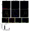
Cigarette Smoke (CS) exposure promotes caspase-1 cleavage in BAL macrophages and neutrophils in an NLRP3-dependent manner. Cleaved caspase-1 immunostaining on BAL cells collected after Air or CS exposure of WT, MCC950 WT treated mice and Nlrp3-/- mice. Cleaved caspase-1 is shown in red and nucleus in blue (DAPI) (A). BAL cells fluorescence intensity quantification (B). Data are representative of three experiments. Bar graph is expressed as mean ± SEM (n =2–4 mice per group, *p < 0.05 ns, non significant, using a Mann Whitney test).
Figure 6
Figure 6
Cigarette Smoke (CS) exposure induces GSDMD cleavage in bronchial epithelial cells in an NLRP3-dependent manner. Immunoblot for total and cleaved GSDMD proteins in lung homogenates of Air or CS-exposed mice of WT, Nlrp3-/- or Gsdmd-/- mice (A) and quantification of total GSDMD (B) and cleaved GSDMD (C) immunoblot were shown. Cleaved GSDMD immunostaining was performed on lung sections of Air or CS-exposed WT, Nlrp3-/- and Gsdmd-/- mice (D). Data are representative of three experiments. Bar graph are expressed ± SEM (n =2–4 mice per group).
Figure 7
Figure 7
Cigarette Smoke (CS) exposure promotes GSDMD expression and cleavage in BAL alveolar macrophages dependently of NLRP3. Cleaved GSDMD immunostaining on BAL cells collected after Air or CS exposure in WT, MCC950-treated WT mice, Nlrp3-/- and Gsdmd-/- mice. Cleaved GSDMD is shown in red and nucleus in blue (DAPI) (A). Airway macrophages fluorescence intensity quantification (B). Data are representative of three experiments. Bar graph is expressed as mean ± SEM (n =2–4 mice per group, *p < 0.05 using a Mann Whitney test).
Figure 8
Figure 8
Gasdermin d-deficient mice (Gsdmd-/- ) have reduced pulmonary inflammation and remodeling after acute smoke cigarette (CS) exposure. WT and Gsdmd-/- mice were exposed to CS or Air during 4 days. Total cells (A), macrophages (B) and neutrophils (C) counts and MPO level in BAL (D) are shown. CXCL1 (E), CXCL5 (F), CXCL15 (G) and IL-1β (H) levels were measured in BAL. MMP-9 (I) and TIMP-1 (J) remodeling factor levels were measured in BAL. In lungs, levels of IL-1β (K), MMP-9 (L) and TIMP-1 (M) were also measured. All parameters decreased in Gsdmd-/- mice exposed to CS as compared to CS WT mice. Data are representative of four experiments and are expressed as mean values ± SEM (n= 4-6 mice per group, *p < 0.05, **p < 0.01, ns, non significant, using a Mann Whitney test).

Similar articles

Cited by

References

    1. Barnes PJ. Cellular and molecular mechanisms of chronic obstructive pulmonary disease. Clin Chest Med (2014) 35:71–86. doi: 10.1016/j.ccm.2013.10.004 - DOI - PubMed
    1. Barnes PJ. Mediators of chronic obstructive pulmonary disease. Pharmacol Rev (2004) 56:515–48. doi: 10.1124/pr.56.4.2 - DOI - PubMed
    1. Barnes PJ, Burney PG, Silverman EK, Celli BR, Vestbo J, Wedzicha JA, et al. . Chronic obstructive pulmonary disease. Nat Rev Dis Primers (2015) 1:15076. doi: 10.1038/nrdp.2015.76 - DOI - PubMed
    1. Wedzicha JA, Singh R, Mackay AJ. Acute COPD exacerbations. Clin Chest Med (2014) 35:157–63. doi: 10.1016/j.ccm.2013.11.001 - DOI - PubMed
    1. Barnes PJ. New anti-inflammatory targets for chronic obstructive pulmonary disease. Nat Rev Drug Discovery (2013) 12:543–59. doi: 10.1038/nrd4025 - DOI - PubMed

Publication types

MeSH terms