

Dach1 transcription factor regulates the expression of peripheral node addressin and lymphocyte trafficking in lymph nodes
- PMID: 36045707
- PMCID: PMC9421177
- DOI: 10.1016/j.crimmu.2022.08.008
Dach1 transcription factor regulates the expression of peripheral node addressin and lymphocyte trafficking in lymph nodes
Abstract
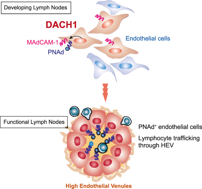
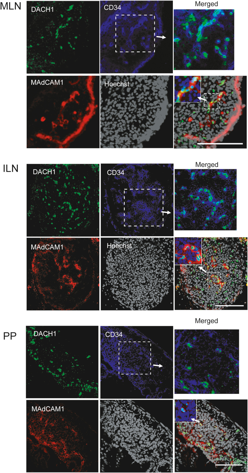
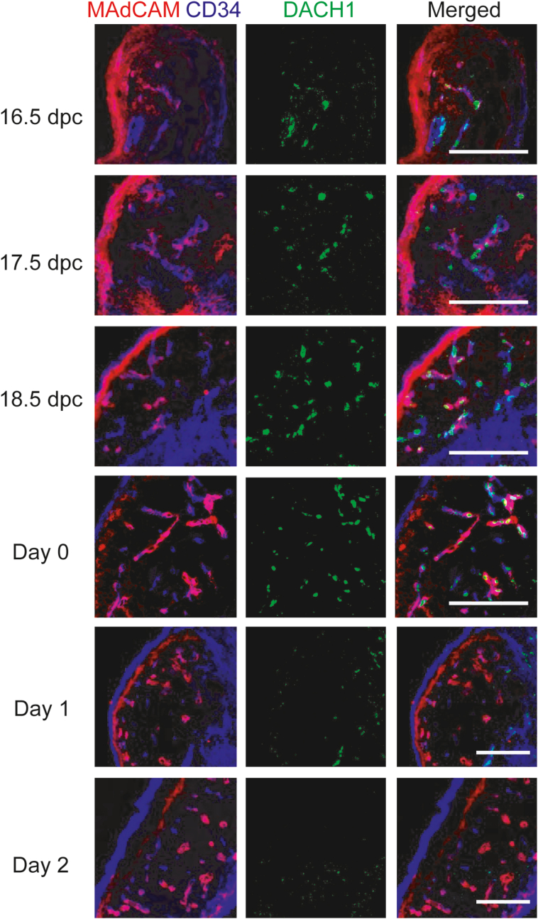
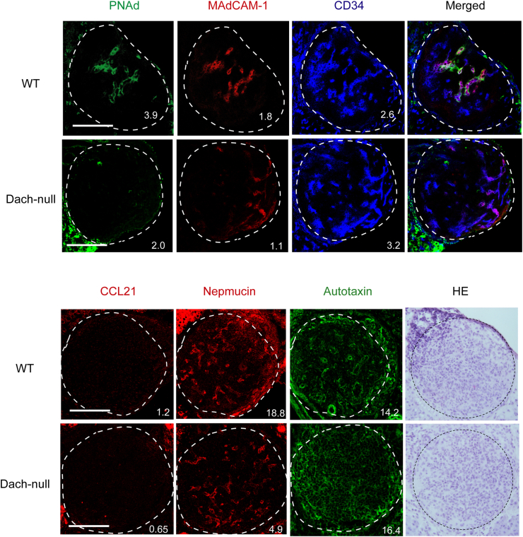
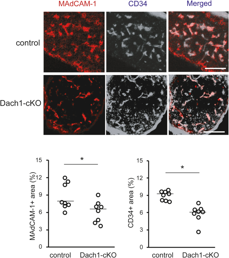
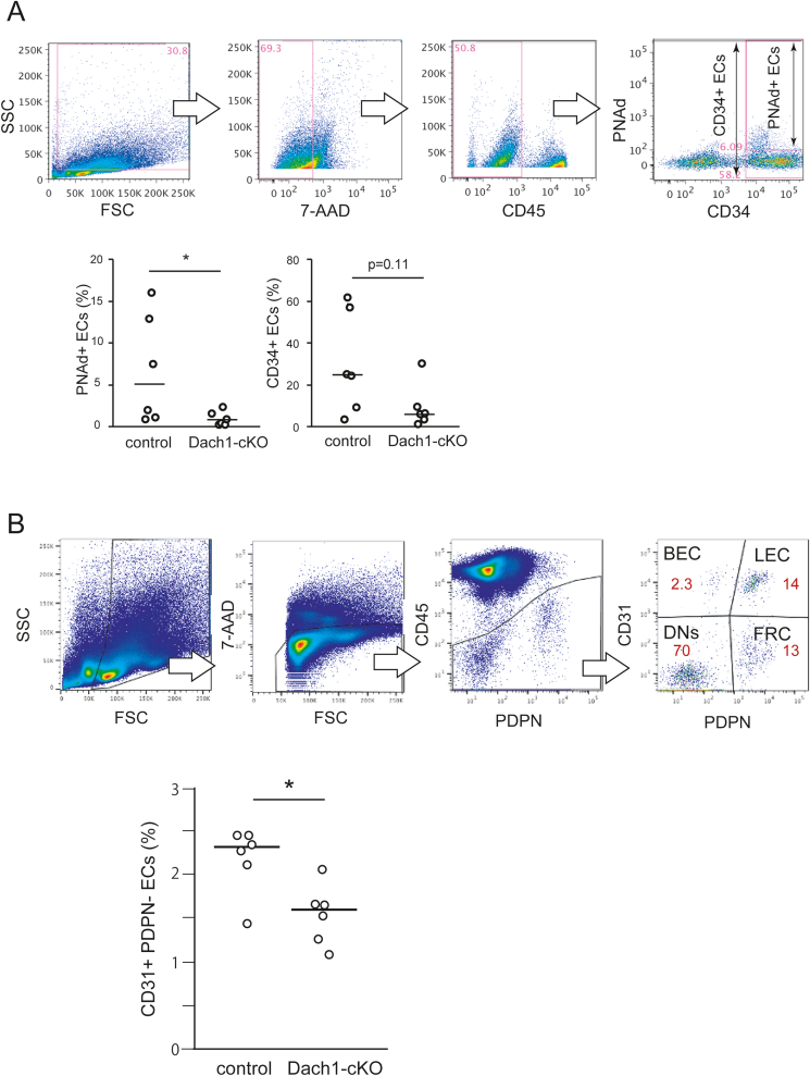
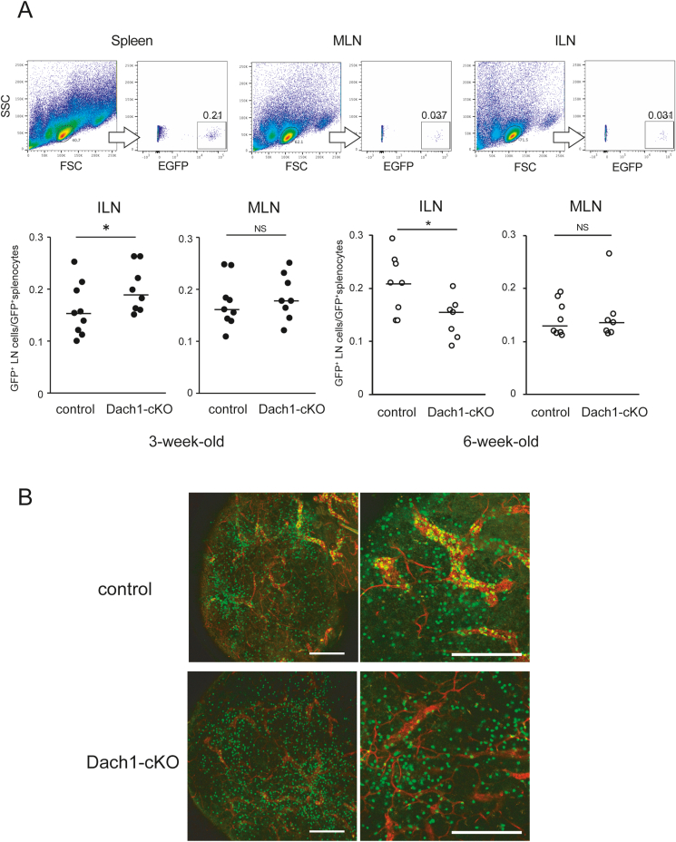
Lymphocytes regulate the immune response by circulating between the vascular and lymphatic systems. High endothelial venules, HEVs, special blood vessels expressing selective adhesion molecules, such as PNAd and MAdCAM-1, mediate naïve lymphocyte migration from the vasculature into the lymph nodes and Peyer's patches. We have identified that DACH1 is abundantly expressed in developing HEV-type endothelial cells. DACH1 showed a restricted expression pattern in lymph node blood vessels during the late fetal and early neonatal periods, corresponding to HEV development. The proportion of MAdCAM-1+ and CD34+ endothelial cells is reduced in the lymph nodes of neonatal conventional and vascular-specific Dach1-deficient mice. Dach1-deficient lymph nodes in adult mice demonstrated a lower proportion of PNAd+ cells and lower recruitment of intravenously administered lymphocytes from GFP transgenic mice. These findings suggest that DACH1 promotes the expression of HEV-selective adhesion molecules and mediates lymphocyte trafficking across HEVs into lymph nodes.
Keywords: ECs, endothelial cells; Endothelial cell; HEV, High endothelial venules; ILNs, Inguinal lymph nodes; LNs, Lymph node; Lymph node; Lymphocyte; MAdCAM-1, mucosal addressin cell adhesion molecule-1; MLNs, Mesenteric lymph nodes; PNAd, peripheral node addressin; PPs, Peyer's patches; Trafficking; Transcription factor; mAb, monoclonal antibody.
© 2022 The Authors.
Conflict of interest statement
The authors declare that they have no known competing financial interests or personal relationships that could have appeared to influence the work reported in this paper.
Figures
Similar articles
-
Absence of Nkx2-3 homeodomain transcription factor reprograms the endothelial addressin preference for lymphocyte homing in Peyer's patches.J Immunol. 2014 Nov 15;193(10):5284-93. doi: 10.4049/jimmunol.1402016. Epub 2014 Oct 15. J Immunol. 2014. PMID: 25320278
-
Nasal-associated lymphoid tissue: phenotypic and functional evidence for the primary role of peripheral node addressin in naive lymphocyte adhesion to high endothelial venules in a mucosal site.J Immunol. 1999 Aug 1;163(3):1382-9. J Immunol. 1999. PMID: 10415038
-
Immunohistologic and functional characterization of a vascular addressin involved in lymphocyte homing into peripheral lymph nodes.J Cell Biol. 1988 Nov;107(5):1853-62. doi: 10.1083/jcb.107.5.1853. J Cell Biol. 1988. PMID: 2460470 Free PMC article.
-
Understanding high endothelial venules: Lessons for cancer immunology.Oncoimmunology. 2015 May 7;4(6):e1008791. doi: 10.1080/2162402X.2015.1008791. eCollection 2015 Jun. Oncoimmunology. 2015. PMID: 26155419 Free PMC article. Review.
-
High Endothelial Venules and Other Blood Vessels: Critical Regulators of Lymphoid Organ Development and Function.Front Immunol. 2017 Feb 3;8:45. doi: 10.3389/fimmu.2017.00045. eCollection 2017. Front Immunol. 2017. PMID: 28217126 Free PMC article. Review.
Cited by
-
Regulation, Maintenance, and Remodeling of High Endothelial Venules in Homeostasis, Inflammation, and Cancer.Curr Opin Physiol. 2023 Dec;36:100705. doi: 10.1016/j.cophys.2023.100705. Epub 2023 Aug 18. Curr Opin Physiol. 2023. PMID: 38523879 Free PMC article.
-
Dach1 is essential for maintaining normal mature podocytes.PLoS One. 2024 May 28;19(5):e0303910. doi: 10.1371/journal.pone.0303910. eCollection 2024. PLoS One. 2024. PMID: 38805434 Free PMC article.
-
The Impact of Mast Cells on the Anatomy, Cellular Communication, and Molecular Immune Network of Lymph Nodes.Clin Rev Allergy Immunol. 2025 Apr 2;68(1):35. doi: 10.1007/s12016-025-09050-5. Clin Rev Allergy Immunol. 2025. PMID: 40175843 Free PMC article. Review.
-
Novel immunologic mechanisms for Fontan-associated liver disease.Int J Cardiol Congenit Heart Dis. 2024 Nov 27;19:100554. doi: 10.1016/j.ijcchd.2024.100554. eCollection 2025 Mar. Int J Cardiol Congenit Heart Dis. 2024. PMID: 39926126 Free PMC article.
-
Epigenetic targets to enhance antitumor immune response through the induction of tertiary lymphoid structures.Front Immunol. 2024 Jan 25;15:1348156. doi: 10.3389/fimmu.2024.1348156. eCollection 2024. Front Immunol. 2024. PMID: 38333212 Free PMC article. Review.
References
-
- Backman M., Machon O., Van Den Bout C.J., Krauss S. Targeted disruption of mouse Dach1 results in postnatal lethality. Dev. Dynam. 2003;226(1):139–144. - PubMed
-
- Baumheter S., Singer M.S., Henzel W., Hemmerich S., Renz M., Rosen S.D., et al. Binding of L-selectin to the vascular sialomucin CD34. Science. 1993;262(5132):436–438. - PubMed
-
- Berg E.L., McEvoy L.M., Berlin C., Bargatze R.F., Butcher E.C. L-selectin-mediated lymphocyte rolling on MAdCAM-1. Nature. 1993;366(6456):695–698. - PubMed
-
- Caubit X., Thangarajah R., Theil T., Wirth J., Nothwang H.G., Ruther U., et al. Mouse Dac, a novel nuclear factor with homology to Drosophila dachshund shows a dynamic expression in the neural crest, the eye, the neocortex, and the limb bud. Dev. Dynam. 1999;214(1):66–80. - PubMed
LinkOut - more resources
Full Text Sources
Molecular Biology Databases
