. 2022 Jul 11:80:104129.
doi: 10.1016/j.amsu.2022.104129. eCollection 2022 Aug.

Effect of intra-peritoneal induction of ascites fluid on the rate of postoperative intraabdominal adhesion in a rat model

Affiliations

Effect of intra-peritoneal induction of ascites fluid on the rate of postoperative intraabdominal adhesion in a rat model

Zahra Amirian et al. Ann Med Surg (Lond). .

Abstract

Introduction: Intra-abdominal adhesions (IAAs) are secondary to peritoneal injuries such as previous surgery or intra-abdominal infections (IAIs). Accordingly, it is crucial to employ fitting techniques to minimize the likelihood of adhesions in any surgery. Due to a paucity of similar data available, this study sought to explore the effects of induced high serum ascites albumin gradient (SAAG) and low serum ascites albumin gradient (SAAG) on the rate of post-operative microscopic and macroscopic adhesion in a mouse model.

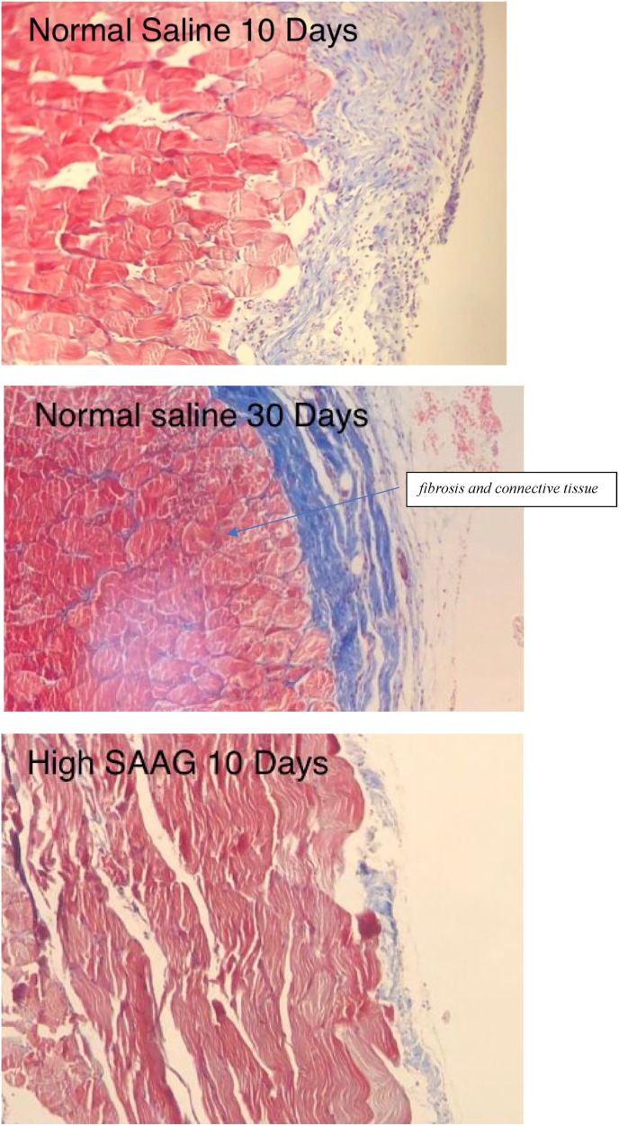
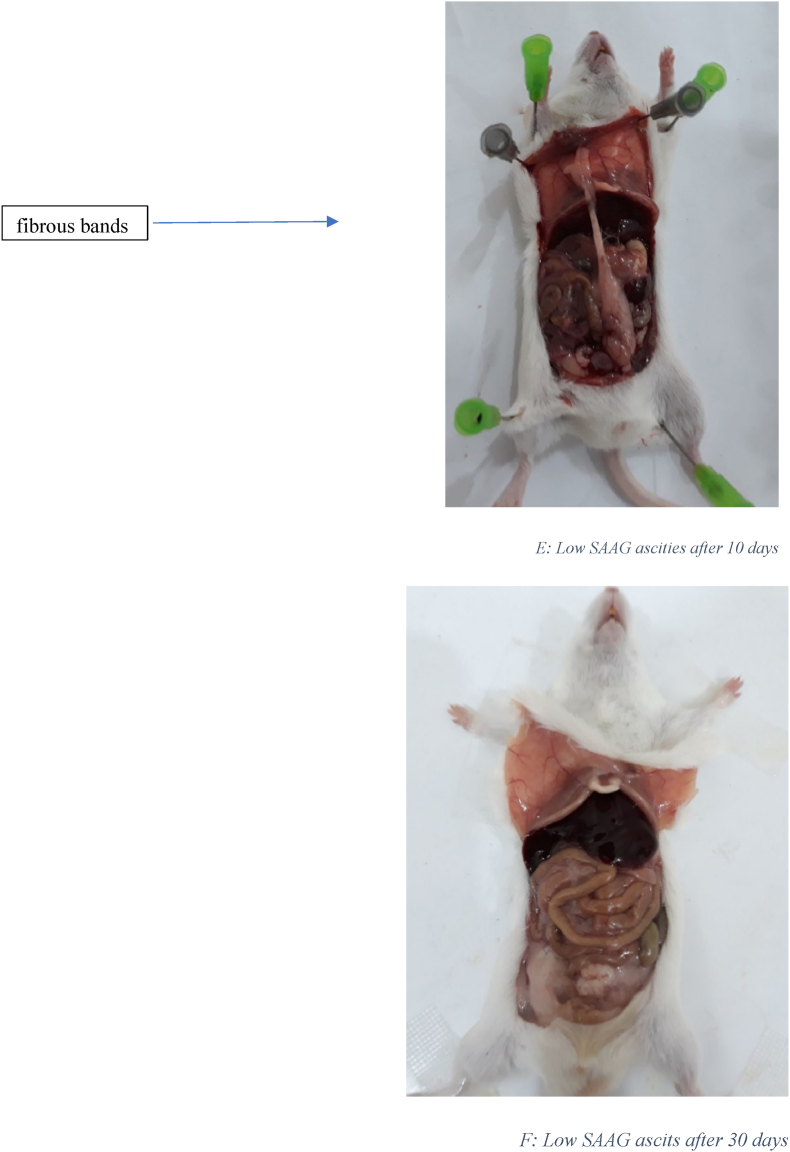
Material and methods: Sixty mice were compared in six groups of ten each. Control groups (1 &4) received normal saline, groups 2&5 received high SAAG ascites fluid, and groups 3&6 received low SAAG ascites fluid intraperitoneally. These groups underwent exploratory laparotomy on day zero, followed by the same procedure on the 10th (groups 1,2,3) and the 30th (Groups 4,5,6) day of surgery. Then, microscopic and macroscopic IAAs were evaluated. Data were analyzed in SPSS software and compared with a p-value less than 0.05.

Results: By comparison, the least microscopic and macroscopic IAAs after 10 and 30 days were found in the low SAAG ascites group. Revealing a statistically significant difference compared to the other two groups (P = 0.01). After 10 days of surgery, macroscopic IAA in the high SAAG group was significantly lower compared to the control and Low SAAG ascites groups.

Conclusion: Intraabdominal low SAAG ascites fluid can significantly decrease the probability of postoperative fibrosis and adhesion band formation.

Protocol number: IR. BUMS.REC.1399.503.

Keywords: Ascites fluid; High SAAG; Intra-abdominal adhesions (IAA); Low SAAG.


Conflict of interest statement

The authors declare no competing interest.

Figures

Fig. 1
Fig. 1
Masson's trichrome stain. Specifically, to distinguish cells from surrounding connective tissue. Connective tissue is stained blue, nuclei are stained dark red/purple, and cytoplasm is stained red/pink. (For interpretation of the references to colour in this figure legend, the reader is referred to the Web version of this article.)
Fig. 1
Fig. 1
Masson's trichrome stain. Specifically, to distinguish cells from surrounding connective tissue. Connective tissue is stained blue, nuclei are stained dark red/purple, and cytoplasm is stained red/pink. (For interpretation of the references to colour in this figure legend, the reader is referred to the Web version of this article.)
Fig. 2
Fig. 2
A: Intra-abdominal view10 days after laparotomy in the normal salin group: 8 rats had 3 or more than 3 fibrous bands B: Intra-abdominal view 30 days after laparotomy in the normal salin group: 9 rats had more than 3 fibrous bands C: Intra-abdominal view10 days after laparotomy in High SAAG ascits group: 5 rats had 3 or more than 3 fibrous bands D: Intra-abdominal view 30 days after laparotomy in High SAAG ascits group: 6 rats had 3 or more than 3 fibrous band E: Intra-abdominal view10 days after laparotomy in Low SAAG ascits group: 2 rats had 3 fibrous bands, no more than 3 fibrous bands were seen, some adhesion were seen F: Intra-abdominal view 30 days after laparotomy in Low SAAG ascits group: 2 rats had 3 fibrous bands, no more than 3 fibrous bands were seen, No adhesion.
Fig. 2
Fig. 2
A: Intra-abdominal view10 days after laparotomy in the normal salin group: 8 rats had 3 or more than 3 fibrous bands B: Intra-abdominal view 30 days after laparotomy in the normal salin group: 9 rats had more than 3 fibrous bands C: Intra-abdominal view10 days after laparotomy in High SAAG ascits group: 5 rats had 3 or more than 3 fibrous bands D: Intra-abdominal view 30 days after laparotomy in High SAAG ascits group: 6 rats had 3 or more than 3 fibrous band E: Intra-abdominal view10 days after laparotomy in Low SAAG ascits group: 2 rats had 3 fibrous bands, no more than 3 fibrous bands were seen, some adhesion were seen F: Intra-abdominal view 30 days after laparotomy in Low SAAG ascits group: 2 rats had 3 fibrous bands, no more than 3 fibrous bands were seen, No adhesion.

References

    1. Bunicard F.C.A.D., Bihhiav T.R., Dunn D.L., Hunter J.G., Mathews J.B., Pollack . The McGraw-Hill Company; 2015. Schwartz Principles of Surgery 2015. Schwartz Principles of Surgery 2015; p. 224.
    1. Tingstedt B., Isaksson K., Andersson E., Andersson R. Prevention of abdominal adhesions–present state and what's beyond the horizon? Eur. Surg. Res. 2007;39(5):259–268. - PubMed
    1. Okabayashi K., Ashrafian H., Zacharakis E., Hasegawa H., Kitagawa Y., Athanasiou T., et al. Adhesions after abdominal surgery: a systematic review of the incidence, distribution and severity. Surg. Today. 2014;44(3):405–420. - PubMed
    1. Wilkins M., Indar A., Wormald R. Intraoperative mitomycin C for glaucoma surgery. Cochrane Database Syst. Rev. 2005;(4) - PMC - PubMed
    1. Penzias A., Bendikson K., Falcone T., Gitlin S., Gracia C., Hansen K., et al. Postoperative adhesions in gynecologic surgery: a committee opinion. Fertil. Steril. 2019;112(3):458–463. - PubMed