Case Reports
. 2022 Jul 25;4(4):CASE22184.
doi: 10.3171/CASE22184.

Spinal intraosseous arteriovenous fistulas with perimedullary drainage associated with vertebral compression fracture: illustrative case

Affiliations
Case Reports

Spinal intraosseous arteriovenous fistulas with perimedullary drainage associated with vertebral compression fracture: illustrative case

Hiroshi Baba et al. J Neurosurg Case Lessons. .

Abstract

Background: Although osseous involvement is occasionally observed in spinal epidural arteriovenous fistulas (AVFs) or seen as a part of diseases of spinal arteriovenous metameric syndrome, purely intraosseous spinal AVFs are extremely rare. Their clinical and imaging characteristic features are not well known. The authors present a case of purely intraosseous AVFs associated with compression fracture.

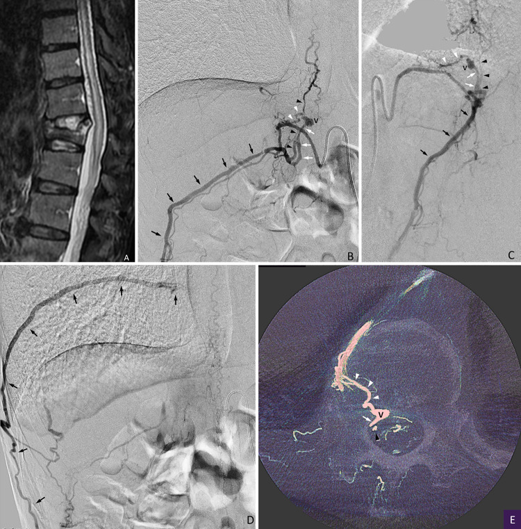
Observations: A 76-year-old man presented with back pain and progressive myelopathy. Computed tomography showed compression fracture of the T12 vertebral body and dilatation of perimedullary veins. Spinal angiography revealed an intraosseous AVF at the T12 spine level, which was fed by multiple feeders of ventral somatic branches and drained into the paravertebral and perimedullary veins. The intraosseous AVF was completely occluded by the combined techniques of transarterial and transvenous embolization with glue and a coil. The symptoms disappeared within 1 month after embolization.

Lessons: Although extremely rare, spinal intraosseous AVFs can develop after compression fracture and cause congestive myelopathy. Combined transarterial and transvenous embolization is useful for the specific case of spinal intraosseous AVFs with both paravertebral and perimedullary drainage.

Keywords: AVF = arteriovenous fistula; CT = computed tomography; MRI = magnetic resonance imaging; NBCA = n-butyl cyanoacrylate; compression fracture; embolization; spinal arteriovenous fistula; spinal arteriovenous malformation; spinal artery; spinal vein.


Conflict of interest statement

Disclosures The authors report no conflict of interest concerning the materials or methods used in this study or the findings specified in this paper.

Figures

FIG. 1.
FIG. 1.
Sagittal fat-suppressed T2-weighted image (A) shows the high signal intensity of the spinal cord and conus medullaris with multiple small flow voids surrounding the spinal cord. A compression fracture of the T12 vertebral body is also noted. Frontal views (B and D), lateral view (C), and axial reformatted image (E) of selective angiography of the right subcostal artery at early (B and C) and late (D) arterial phases show the AVFs fed by multiple ventral somatic branches and draining into the basivertebral vein (white arrowheads) and epidural vein (white arrows) with a varix (V) and then into the paravertebral veins (black arrows) (the subcostal vein, intersegmental anastomotic vein, T10 intercostal vein, and the azygos vein) and retrogradely into the perimedullary veins via the radiculomedullary vein (black arrowheads).
FIG. 2.
FIG. 2.
A: Lateral view of selective angiography of the feeder of a ventral somatic branch shows an AVF (asterisk) draining into the basivertebral vein (white arrowheads), ventral epidural vein (VEV), subcostal vein (SCV), and radiculomedullary vein (black arrowheads). The arrow indicates the tip of a 1.3-Fr microcatheter. B: Lateral view of selective venography shows retrograde intradural drainage via the radiculomedullary vein. The tip of a 1.6-Fr microcatheter (arrowhead) is introduced transvenously into the outlet to the radiculomedullary vein via the subcostal vein (arrows). C: Schematic of the angioarchitecture and embolization techniques of the presented case. The AVFs were fed by multiple ventral somatic branches of the right subcostal artery (R T12 SA) and drained into the basivertebral vein and ventral epidural vein (VEV), forming a varix (V) and then into the subcostal vein (SCV) and the radiculomedullary vein (RMV). A 1.3-Fr microcatheter (1.3F MC) was advanced into a feeder of the ventral somatic branch of the right subcostal artery, and a microballoon catheter was placed in the stem of the subcostal artery. A 4-Fr guiding sheath (4F GS) was placed in the azygos vein (AZV) with a right cubital venous approach, a 1.6-Fr (1.6F MC)/2.6-Fr (2.6F MC) coaxial microcatheter system was introduced into the right subcostal vein (SCV) via the right T10 intercostal vein and a longitudinal anastomotic vein, and a 1.6-Fr microcatheter was further advanced into the extradural portion of the radiculomedullary vein (RMV). D: Lateral view of angiography during injection of 33% NBCA-lipiodol mixture via the 1.6-Fr microcatheter in the extradural portion of the radiculomedullary vein. The glue cast fills the radiculomedullary vein and the epidural vein while partially filling the paravertebral vein. E: Lateral view of angiography during injection of 20% NBCA-lipiodol mixture via the 1.3-Fr microcatheter in the feeder. The glue cast fills beyond the fistula into the basivertebral vein and the epidural varix. F: Axial reformatted image of rotational angiography without contrast injection clearly shows the glue cast in the feeder (a ventral somatic branch), the basivertebral vein, and the epidural varix (V). The arrow indicates the radiculomedullary vein embolized transvenously with glue and coil.
FIG. 3.
FIG. 3.
Frontal view of selective angiography of the right subcostal artery immediately after embolization shows disappearance of the AVFs.

References

    1. Rangel-Castilla L, Holman PJ, Krishna C, Trask TW, Klucznik RP, Diaz OM. Spinal extradural arteriovenous fistulas: a clinical and radiological description of different types and their novel treatment with Onyx. J Neurosurg Spine. 2011;15(5):541–549. - PubMed
    1. Kiyosue H, Matsumaru Y, Niimi Y, et al. Angiographic and clinical characteristics of thoracolumbar spinal epidural and dural arteriovenous fistulas. Stroke. 2017;48(12):3215–3222. - PMC - PubMed
    1. Mull M, Othman A, Dafotakis M, Hans FJ, Schubert GA, Jablawi F. Spinal epidural arteriovenous fistula with perimedullary venous reflux: clinical and neuroradiologic features of an underestimated vascular disorder. AJNR Am J Neuroradiol. 2018;39(11):2095–2102. - PMC - PubMed
    1. Wendl CM, Aguilar Pérez M, Felber S, Stroszczynski C, Bäzner H, Henkes H. Paraspinal arteriovenous fistula: Stuttgart classification based on experience and a review of the literature. Br J Radiol. 2018;91(1088):20170337. - PMC - PubMed
    1. Chul Suh D, Gon Choi C, Bo Sung K, Kim KK, Chul Rhim S. Spinal osseous epidural arteriovenous fistula with multiple small arterial feeders converging to a round fistular nidus as a target of venous approach. AJNR Am J Neuroradiol. 2004;25(1):69–73. - PMC - PubMed

Publication types

LinkOut - more resources