Review
. 2022 Aug 15:10:931311.
doi: 10.3389/fcell.2022.931311. eCollection 2022.

Evolution of astrocytes: From invertebrates to vertebrates

Affiliations
Review

Evolution of astrocytes: From invertebrates to vertebrates

Carmen Falcone. Front Cell Dev Biol. .

Abstract

The central nervous system (CNS) shows incredible diversity across evolution at the anatomical, cellular, molecular, and functional levels. Over the past decades, neuronal cell number and heterogeneity, together with differences in the number and types of neuro-active substances, axonal conduction, velocity, and modes of synaptic transmission, have been rigorously investigated in comparative neuroscience studies. However, astrocytes, a specific type of glial cell in the CNS, play pivotal roles in regulating these features and thus are crucial for the brain's development and evolution. While special attention has been paid to mammalian astrocytes, we still do not have a clear definition of what an astrocyte is from a broader evolutionary perspective, and there are very few studies on astroglia-like structures across all vertebrates. Here, I elucidate what we know thus far about astrocytes and astrocyte-like cells across vertebrates. This information expands our understanding of how astrocytes evolved to become more complex and extremely specialized cells in mammals and how they are relevant to the structure and function of the vertebrate brain.

Keywords: astrocyte; astrocytes; central nervous system; evolution; glia; vertebrates.


Conflict of interest statement

The authors declare that the research was conducted in the absence of any commercial or financial relationships that could be construed as a potential conflict of interest.

Figures
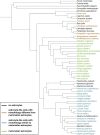
FIGURE 1
FIGURE 1
Evolutionary tree of the species discussed in this study. The cladogram was created on https://phylot.biobyte.de/, based on the NCBI taxonomy database. Color code: black = no astrocytes; orange = species showing astrocyte-like cells with morphology different from mammalian astrocytes; blue = species showing astrocyte-like cells with morphology similar to mammalian astrocytes; green = species showing mammalian astrocyte morphology.
FIGURE 2
FIGURE 2
Schematic of cell types considered as predecessors of mammalian astrocytes. (A) Ependymal cells. (B) Radial glia cells. (C) Astrocyte-like cells (e.g. as seen in reptiles).
FIGURE 3
FIGURE 3
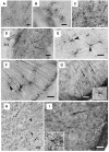
Astrocyte-like cells in reptile brains. (A) Adapted from Lõrincz and Kálmán, 2020, from Figure 4H. GFAP-immunopositive elements in agama telencephalon; arrows point to astrocytes intermingled within RG processes in the nucleus accumbens. Scale bar: 20 µm. (B) Adapted from Lõrincz and Kálmán, 2020, from Figure 5F. Astrocytes from the chameleon septum. Scale bar: 20 µm. (C) Adapted from Lõrincz and Kálmán, 2020, from Figure 5G. Astrocytes from the chameleon hypothalamus. Scale bar: 20 µm. (D) Adapted from Lõrincz and Kálmán, 2020, from Figure 8C. Arrows point to astrocytes within the optic tract (TO) of Moroccan eyed lizard diencephalon. Scale bar: 80 µm. (E) Adapted from Lõrincz and Kálmán, 2020, from Figure 9J. Arrowheads point to astrocytes in the chameleon brain. Scale bar: 20 µm. (F) Arrowheads point to astrocytes soma in the chameleon brain. Scale bar: 20 µm. (G) Adapted from Lõrincz and Kálmán, 2020, from Figure 13J. Arrows point to astrocytes with long processes in the brain stem of the python. Scale bar: 50 µm. (H) Adapted from Lõrincz and Kálmán, 2020, from Figure 10G. Astrocytes in the corn snake brain; arrows point to astrocytes, arrowheads point to RG processes. Scale bar: 40 µm. (I) Adapted from Lõrincz and Kálmán, 2020, from Figure 13L. Astrocytes in the ventrolateral part of the brain stem in the corn snake; arrow points to a cell enlarged in the inset. Scale bar: 50 µm.
FIGURE 4
FIGURE 4
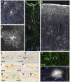
Astrocytes in hominid brains. (A) Adapted from Oberheim et al., 2009, Figure 4B. Typical human protoplasmic astrocyte. White: GFAP; blue: DAPI. Scale bar: 20 µm. (B) Adapted from Oberheim et al., 2009, Figure 7B. Human fibrous astrocytes in white matter. Grey: GFAP. Scale bar: 10 µm. (C) Example of ILA in the rhesus macaque dorsofrontal cortex. Green: GFAP; Red: Lectin; Blue: DAPI. Scale bar: 20 µm. (D) Adapted from Oberheim et al., 2009, Figure 3A, showing ILA palisade. Pial surface and layers one to two of human cortex. Dashed yellow line indicates border between layer 1 and 2. White: GFAP; Blue: DAPI. Scale bar, 100 µm. (E–E”) Adapted from Falcone et al., 2021, Figure 2A. GFAP + VP-A in frontal cortex of a gibbon. Scale bar: 30 μm. (E′,E″) Higher magnification of 1 and 2 in E, respectively. Scale bar = 10 μm. (F,F′) Adapted from Falcone et al., 2021, Figure 2C. GFAP + VP-A in frontal cortex of a gorilla. Scale bar = 30 μm. (F′) Higher magnification of squared area in (F). Scale bar: 10 μm. (G–G″) Adapted from Falcone et al., 2021, Figure 2D. GFAP+ VP-A in the human frontal cortex. Scale bar = 30 μm. (G′,G″) Higher magnification of 1 and 2 in G, respectively. Arrows point to cell somata, arrowheads point to varicosities on VP-A processes. Scale bar: 10 μm. (H) Adapted from Falcone et al., 2021, Figure 3E. VP-A in human frontal cortex. Green = GFAP; Blue = DAPI. Scale bar = 20 μm. Arrows point to cell somata, arrowheads point to varicosities on the VP-A processes. (I,I′) Adapted from Oberheim et al., 2009, Figure 2B. Diolistic labeling (white) of a VP-A whose long process terminates in the neuropil. Blue = sytox. Scale bar: 20 µm. (I′) High-power image of the yellow box in I highlighting the varicosities seen along the processes. Scale bar, 10 µm.

References

    1. Achúcarro N. (1915). De l’évolution de La Névroglie et Spécialement de Ses Relations Avec l’appareil Vasculaire (Avec 24 Gravures). no 13, 169–212.
    1. Andriezen W. L. (1893). The neuroglia elements in the human brain. Br. Med. J. 2 (1700), 227–230. 10.1136/bmj.2.1700.227 - DOI - PMC - PubMed
    1. Bacaj T., Tevlin M., Lu Y., Shaham S. (2008). Glia are essential for sensory organ function in C. Elegans. Sci. (New York, N.Y.) 322 (5902), 744–747. 10.1126/science.1163074 - DOI - PMC - PubMed
    1. Bairati A., Maccagnani F. (1950). Ricerche sulla glioarchitettonica dei vertebrati. II uccelli. Monit. Zool. Ital. 58, 53–55.
    1. Bairati A., Tripoli G. (1954). [Morphology and histochemistry of the glia of neural axis in vertebrates]. Z. Für. Zellforsch. 39 (4), 392–413. 10.1007/BF00343889 - DOI - PubMed