. 2022 Aug 22:2022:1614336.
doi: 10.1155/2022/1614336. eCollection 2022.

Hypoxia-Driven M2-Polarized Macrophages Facilitate Cancer Aggressiveness and Temozolomide Resistance in Glioblastoma

Affiliations

Hypoxia-Driven M2-Polarized Macrophages Facilitate Cancer Aggressiveness and Temozolomide Resistance in Glioblastoma

Ge Zhang et al. Oxid Med Cell Longev. .

Abstract

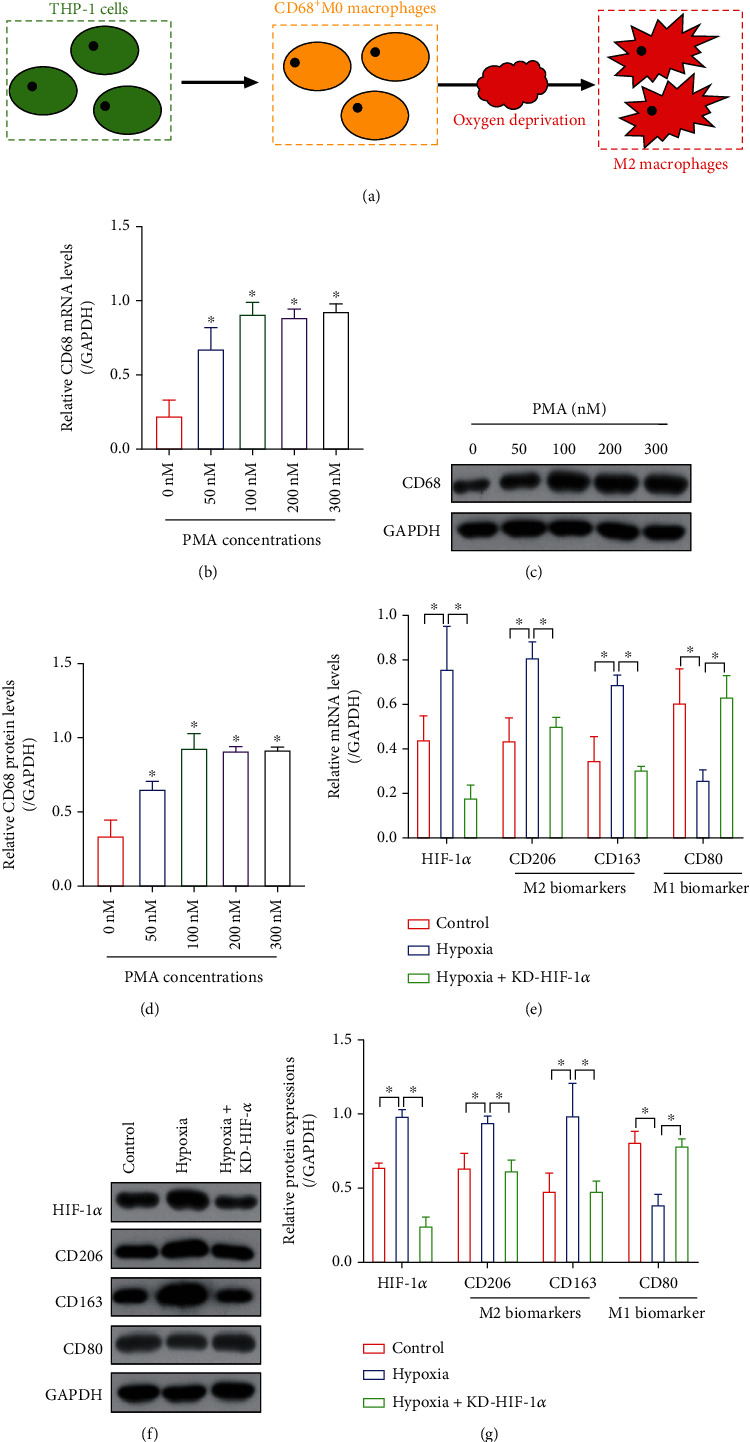
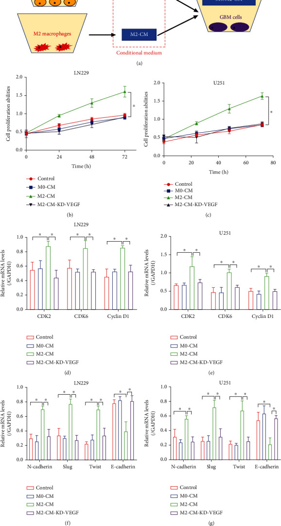
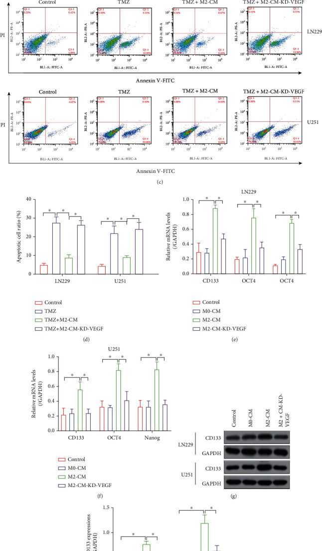
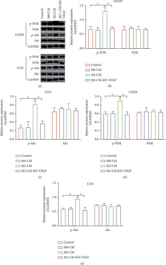
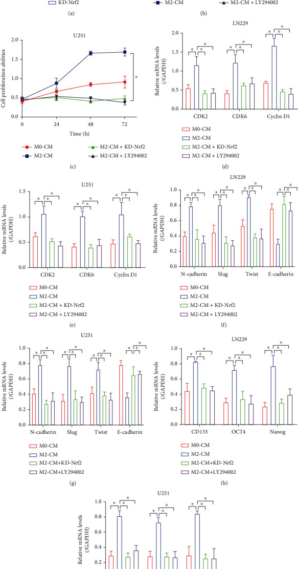
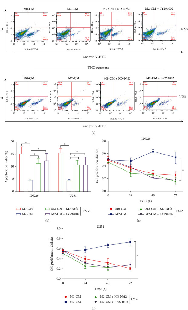
Hypoxia-induced M2 phenotypes of tumor associated macrophages (TAMs) promote the development and chemoresistance of multiple types of cancers, including glioblastoma (GBM). However, the detailed molecular mechanisms have not been fully understood. In this study, we firstly reported that hypoxic pressure promoted M2 macrophage generation, which further promoted cancer progression and temozolomide (TMZ) resistance in GBM through secreting vascular endothelial growth factor (VEGF). Specifically, the clinical data suggested that M2 macrophages were significantly enriched in GBM tissues compared with the adjacent normal tissues, and the following in vitro experiments validated that hypoxic pressure promoted M2-polarized macrophages through upregulating hypoxia-inducible factor-1α (HIF-1α). In addition, hypoxic M2 macrophages VEGF-dependently promoted cell proliferation, epithelial-mesenchymal transition (EMT), glioblastoma stem cell (GSC) properties, and TMZ resistance in GBM cells through activating the PI3K/Akt/Nrf2 pathway. Also, M2 macrophages secreted VEGF to accelerate angiogenesis in human umbilical vein endothelial cells (HUVECs) through interacting with its receptor VEGFR. In general, we concluded that hypoxic M2 macrophages contributed to cancer progression, stemness, drug resistance, and angiogenesis in GBM through secreting VEGF, and our data supported the notion that targeting hypoxia-associated M2 macrophages might be an effective treatment strategy for GBM in clinical practices.


Conflict of interest statement

The authors have no conflict of interest.

Figures

Figure 1
Figure 1
M1/M2 macrophages were closely related with GBM progression, prognosis, and drug resistance in clinic. (a, b) The expression levels of CD206 and CD80 in GBM tissues were examined by Real-Time qPCR analysis. (c, d) Kaplan-Meier survival analysis was performed to analyze the correlation of M1/M2 macrophages with patients' prognosis. The correlations of M1/M2 macrophages with (e, f) tumor volume and (g, h) WHO grade were analyzed. (i, j) The relevance of M1/M2 macrophages with TMZ resistance in the clinical GBM tissues was analyzed by performing Real-Time qPCR analysis. Each experiment repeated at least 3 times, and P < 0.05.
Figure 2
Figure 2
Hypoxic conditions promoted M0-to-M2 transition in the THP-1 cells. (a) The graphical illustration for the induction of M2 macrophages from THP-1 cells. PMA promoted the generation of CD68-positive M0 macrophages in a dose-dependent manner, as it was, respectively, determined by performing (b) Real-Time qPCR and (c, d) Western Blot analysis. (e) Real-Time qPCR and (g, g) Western Blot analysis verified that oxygen deprivation promoted M2 macrophage generation through inducing the upregulation of HIF-1α. Each experiment repeated at least 3 times, and P < 0.05.
Figure 3
Figure 3
Hypoxic M2 macrophage-derived conditional medium facilitates GBM aggressiveness and angiogenesis in a VEGF-dependent manner. (a) The graphical illustration for the coculture of GBM cells and the macrophage-derived conditional medium. (b, c) GBM cell proliferation abilities were determined by performing MTT assay. (d, e) The cell mitosis-associated biomarkers (CDK2, CDK6, and Cyclin D1) were examined by performing Real-Time qPCR analysis. (f, g) The EMT-associated signatures (N-cadherin, Slug, Twist, and E-cadherin) were examined by Real-Time qPCR analysis. Each experiment repeated at least 3 times, and P < 0.05.
Figure 4
Figure 4
M2-CM VEGF-dependently increased TMZ resistance in the GBM cells. (a, b) MTT assay results verified that M2-CM increased cell proliferation in the GBM cells cotreated with TMZ. (c, d) FCM assay verified that M2-CM suppressed TMZ-induced apoptotic cell death in the GBM cells. (e, f) Real-Time qPCR and (g, h) Western Blot analysis were used to examine the expression status of GSC-related proteins (CD133, OCT4, and Nanog). Each experiment repeated at least 3 times, and P < 0.05.
Figure 5
Figure 5
M2-CM activated the PI3K/Akt/Nrf2 pathway in a VEGF-dependent manner. (a–e) The expression levels of p-PI3K, PI3K, p-Akt, and Akt in the GBM cells were determined by performing Western Blot analysis. Each experiment repeated at least 3 times, and P < 0.05.
Figure 6
Figure 6
M2-CM promoted cancer aggressiveness through activating the PI3K/Akt/Nrf2 pathway. (a) The shRNA for Nrf2 was delivered into the GBM cells, and the transfection efficiency was determined by Real-Time qPCR. (b, c) Cell proliferation abilities were determined by performing MTT assay. (d, e) Real-Time qPCR was performed to determine the expression status of CDK2, CDK6, and Cyclin D1. (f, g) The EMT-associated markers were examined by conducting Real-Time qPCR. (h, i) The expression levels of the cancer stem cell biomarkers were determined by Real-Time qPCR analysis. Each experiment repeated at least 3 times, and P < 0.05.
Figure 7
Figure 7
M2-CM increased the TMZ resistance in the GBM cells by regulating the PI3K/Akt/Nrf2 pathway. (a, b) The apoptotic cell ratio in the GBM cells was determined by performing the FCM assay. (c, d) The MTT assay was employed to determine cell proliferation in the GBM cells. Each experiment repeated at least 3 times, and P < 0.05.

References

    1. Jacob F., Salinas R. D., Zhang D. Y., et al. A patient-derived glioblastoma organoid model and biobank recapitulates inter- and intra-tumoral heterogeneity. Cell . 2020;180(1):188–204.e22. doi: 10.1016/j.cell.2019.11.036. - DOI - PMC - PubMed
    1. Lah T. T., Novak M., Breznik B. Brain malignancies: glioblastoma and brain metastases. Seminars in Cancer Biology . 2020;60:262–273. doi: 10.1016/j.semcancer.2019.10.010. - DOI - PubMed
    1. De Leo A., Ugolini A., Veglia F. Myeloid cells in glioblastoma microenvironment. Cells . 2021;10(1):p. 18. doi: 10.3390/cells10010018. - DOI - PMC - PubMed
    1. DeCordova S., Shastri A., Tsolaki A. G., et al. Molecular heterogeneity and immunosuppressive microenvironment in glioblastoma. Frontiers in Immunology . 2020;11:p. 1402. doi: 10.3389/fimmu.2020.01402. - DOI - PMC - PubMed
    1. Li M., Kirtane A. R., Kiyokawa J., et al. Local targeting of NAD(+) salvage pathway alters the immune tumor microenvironment and enhances checkpoint immunotherapy in glioblastoma. Cancer Research . 2020;80(22):5024–5034. doi: 10.1158/0008-5472.CAN-20-1094. - DOI - PMC - PubMed

MeSH terms

Substances