. 2023 Apr;41(2):422-433.
doi: 10.5534/wjmh.220009. Epub 2022 Jul 14.

Analysis of 29 Targeted Genes for Non-Obstructive Azoospermia: The Relationship between Genetic Testing and Testicular Histology

Affiliations

Analysis of 29 Targeted Genes for Non-Obstructive Azoospermia: The Relationship between Genetic Testing and Testicular Histology

Rossella Cannarella et al. World J Mens Health. 2023 Apr.

Abstract

Purpose: To analyze the presence of potentially pathogenic variants of 29 candidate genes known to cause spermatogenic failure (SPGF) in patients with non-obstructive azoospermia (NOA) who underwent testicular histology.

Materials and methods: Forty-eight patients with unexplained NOA referred to the Department of Transfusion Medicine and Transplantation Biology, University Hospital Center Zagreb, Zagreb, Croatia for testicular biopsy. They were divided into three groups: those who had cryptorchidism (n=9), those with varicocele (n=14), and those with idiopathic NOA (n=25). All included patients underwent blood withdrawal for next-generation sequencing (NGS) analysis and gene sequencing.

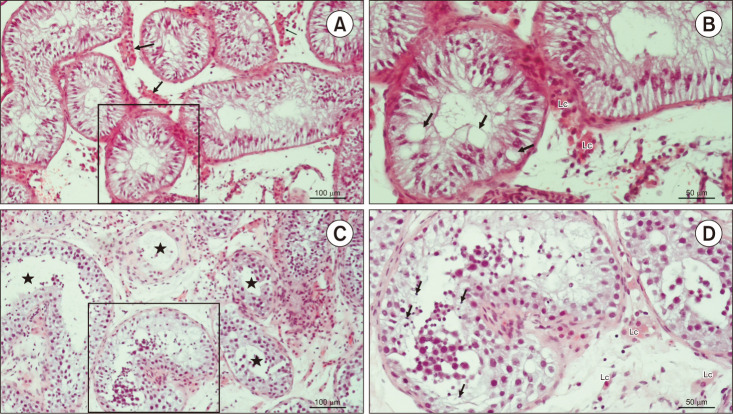
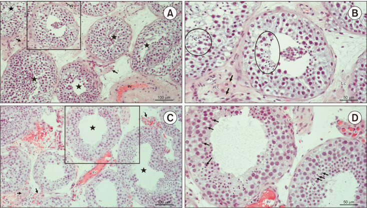
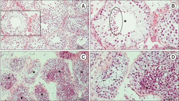
Results: We found a possible genetic cause in 4 patients with idiopathic NOA (16%) and in 2 with cryptorchidism (22%). No pathogenic or possibly pathogenic mutations were identified in patients with varicocele. Variants of undetermined significance (VUS) were found in 11 patients with idiopathic NOA (44%), 3 with cryptorchidism (33%), and 8 patients with varicocele (57%). VUSs of the USP9Y gene were the most frequently as they were found in 14 out of 48 patients (29%). In particular, the VUS USP9Y c.7434+14del was found in 11 patients. They showed varied histological pictures, including Sertoli cell-only syndrome, mixed atrophy, and hypospermatogenesis, regardless of cryptorchidism or varicocele. No direct correlation was found between the gene mutation/variant and the testicular histological picture.

Conclusions: Different mutations of the same gene cause various testicular histological pictures. These results suggest that it is not the gene itself but the type of mutation/variation that determines the testicular histology picture. Based on the data presented above, it remains challenging to design a genetic panel with prognostic value for the outcome of testicular sperm extraction in patients with NOA.

Keywords: Azoospermia; Male infertility; Next-generation sequencing; Spermatogenesis; Testicular histology.


Conflict of interest statement

The authors have nothing to disclose.

Figures

Fig. 1
Fig. 1. Flowchart of the patients included in the study. Out of the 60 patients assessed for eligibility, 48 were finally included. These were divided into 3 groups, those with cryptorchidism, varicocele, and idiopathic NOA. NOA: non-obstructive azoospermia, GCNIS: germ cell neoplasia in situ, CFTR: cystic fibrosis transmembrane conductance regulator.
Fig. 2
Fig. 2. Testicular histology of patients code 160585017 and 141879319. (A) Testicular biopsy from an azoospermic patient. Seminiferous tubules display narrow lumen and are lined with Sertoli cells exclusively. Lc clusters are seen between the seminiferous tubules (→) (H&E, ×200, scale=100 µm). Patient 160585017. (B) Detail from panel A. Due to the depletion of spermatogenic cells, Sertoli cells display giant vacuoles in their cytoplasm (→) (H&E, ×200, scale=50 µm). Patient 160585017. (C) An azoospermic patient with mixed atrophy of seminiferous tubules. Seminiferous epithelium displays a variable status of spermatogenesis (★) (H&E, ×200, scale=100 µm). Patient 141879319. (D) Detail from panel C. In one of the seminiferous tubules, late spermatids (→) are clearly visible, despite the disorganisation of the seminiferous epithelium (H&E, ×200, scale bar=50 µm). Patient 141879319. Lc: Leydig cells.
Fig. 3
Fig. 3. Testicular histology of patients code 265952375 and 119526696. (A) Testicular biopsy from a patient with hypospermatogenesis. In most tubules, (★) seminiferous epithelium is slightly disorganised but bears late spermatids. Within the loose connective tissue, there are clusters of Lc of moderate size (→) (H&E, ×200, scale bar=100 µm). Patient 265952375. (B) Detail from panel A. Despite changed stratification of spermatogenic cells, seminiferous tubules preserved the capacity to produce late spermatids (circled areas). The interstitial compartment bears small BV and Lc. Some Lc are rich in Reinke’s crystals (→) (H&E, ×200, scale bar=50 µm). Patient 265952375. (C) Testicular parenchyma sample from an azoospermic man. Seminiferous tubules display heterogeneous morphology of spermatogenesis (hypospermatogenesis, maturation arrest), including some tubules rich in late spermatids (★). Clusters of Lc (→) have an abundant network of accompanying capillaries. As a consequence of surgery, extravasated erythrocytes (E) are frequently encountered in the bioptic material (H&E, ×200, scale bar=100 µm). Patient 119526696. (D) Detail of panel C. These two seminiferous tubules have maintained a regular diameter and morphology of spermatogenesis. Some late spermatids (→) are in the final stage of spermiogenesis and will be released as spermatozoa in the lumen. Peritubular Lc are found adjacent to the lamina propria of the tubule. The interstitium is rich in BV (H&E, ×200, scale=50 µm). Patient 119526696. Lc: Leydig cells, BV: blood vessels.
Fig. 4
Fig. 4. Testicular histology of patients code 122836561 and 16368087. (A) Testicular biopsy from an azoospermic patient with maturation arrest. The tubules are lined with Sertoli cells and all types of spermatogenic cells, apart from late spermatids (H&E, ×200, scale bar=100 µm). Patient 122836561. (B) Detail of panel A. Seminiferous tubule (★) with the “spermatid stop” (maturation arrest). Spermatogenesis progresses only to the stage of round (early) spermatids (circled area) (H&E, ×200, scale=50 µm). Patient 122836561. (C) A testicular tissue sample from an azoospermic patient with hypospermatogenesis. The stratification of seminiferous epithelium (★) is not maintained. However, all spermatogenic cells, including late spermatids, are visible (H&E, ×200, scale bar=100 µm). Patient 163680875. (D) Detail of panel C. Despite the disorganised seminiferous epithelium, many late spermatids (→) can be identified (H&E, ×200, scale=50 µm). Patient 163680875. Lc: Leydig cells, BV: blood vessels.

References

    1. Punab M, Poolamets O, Paju P, Vihljajev V, Pomm K, Ladva R, et al. Causes of male infertility: a 9-year prospective monocentre study on 1737 patients with reduced total sperm counts. Hum Reprod. 2017;32:18–31. - PMC - PubMed
    1. Tüttelmann F, Ruckert C, Röpke A. Disorders of spermatogenesis: perspectives for novel genetic diagnostics after 20 years of unchanged routine. Med Genet. 2018;30:12–20. - PMC - PubMed
    1. Levine H, Jørgensen N, Martino-Andrade A, Mendiola J, Weksler-Derri D, Mindlis I, et al. Temporal trends in sperm count: a systematic review and meta-regression analysis. Hum Reprod Update. 2017;23:646–659. - PMC - PubMed
    1. Asero P, Calogero AE, Condorelli RA, Mongioi' L, Vicari E, Lanzafame F, et al. Relevance of genetic investigation in male infertility. J Endocrinol Invest. 2014;37:415–427. - PubMed
    1. Cannarella R, Condorelli RA, Duca Y, La Vignera S, Calogero AE. New insights into the genetics of spermatogenic failure: a review of the literature. Hum Genet. 2019;138:125–140. - PubMed