Review
. 2023 Mar;38(7-9):496-528.
doi: 10.1089/ars.2021.0263. Epub 2022 Nov 1.

KRIT1: A Traffic Warden at the Busy Crossroads Between Redox Signaling and the Pathogenesis of Cerebral Cavernous Malformation Disease

Affiliations
Review

KRIT1: A Traffic Warden at the Busy Crossroads Between Redox Signaling and the Pathogenesis of Cerebral Cavernous Malformation Disease

Andrea Perrelli et al. Antioxid Redox Signal. 2023 Mar.

Abstract

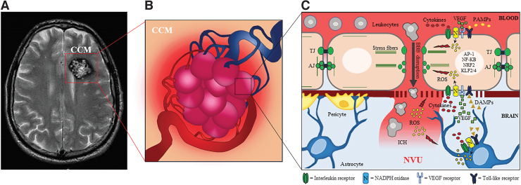
Significance: KRIT1 (Krev interaction trapped 1) is a scaffolding protein that plays a critical role in vascular morphogenesis and homeostasis. Its loss-of-function has been unequivocally associated with the pathogenesis of Cerebral Cavernous Malformation (CCM), a major cerebrovascular disease of genetic origin characterized by defective endothelial cell-cell adhesion and ensuing structural alterations and hyperpermeability in brain capillaries. KRIT1 contributes to the maintenance of endothelial barrier function by stabilizing the integrity of adherens junctions and inhibiting the formation of actin stress fibers. Recent Advances: Among the multiple regulatory mechanisms proposed so far, significant evidence accumulated over the past decade has clearly shown that the role of KRIT1 in the stability of endothelial barriers, including the blood-brain barrier, is largely based on its involvement in the complex machinery governing cellular redox homeostasis and responses to oxidative stress and inflammation. KRIT1 loss-of-function has, indeed, been demonstrated to cause an impairment of major redox-sensitive mechanisms involved in spatiotemporal regulation of cell adhesion and signaling, which ultimately leads to decreased cell-cell junction stability and enhanced sensitivity to oxidative stress and inflammation. Critical Issues: This review explores the redox mechanisms that influence endothelial cell adhesion and barrier function, focusing on the role of KRIT1 in such mechanisms. We propose that this supports a novel model wherein redox signaling forms the common link between the various pathogenetic mechanisms and therapeutic approaches hitherto associated with CCM disease. Future Directions: A comprehensive characterization of the role of KRIT1 in redox control of endothelial barrier physiology and defense against oxy-inflammatory insults will provide valuable insights into the development of precision medicine strategies. Antioxid. Redox Signal. 38, 496-528.

Keywords: GTPases; KRIT1; MAPKs; NADPH oxidases; cerebral cavernous malformation (CCM); cerebrovascular disease; endothelial cell adhesion and barrier function; endothelial-to-mesenchymal transition (EndMT); inflammation; oxidative post-translational modifications (Ox-PTMs); oxidative stress; reactive oxygen species (ROS); redox homeostasis and signaling.


Conflict of interest statement

The authors declare no conflict of interest.

Figures

FIG. 1.
FIG. 1.
CCM. (A) Axial T2-weighted FSE MRI of a CCM in the left cerebral hemisphere of an affected patient, showing a typical mulberry-like internal structure and a surrounding hemosiderin ring (image courtesy of Dr. Maria Consuelo Valentini, “Città della Salute e della Scienza” University Hospital of Torino, Italy). (B) Graphical representation of a typical CCM lesion, which consists of clusters of abnormally enlarged and leaky capillary channels (caverns) having a mulberry-like appearance. (C) In the brain, CCM lesions develop as a result of structural and functional alterations of the NVU, and they may eventually lead to oxy-inflammatory responses, BBB disruption, and ICH. AJ, adherens junction; BBB, blood-brain barrier; CCM, Cerebral Cavernous Malformation; DAMPs, damage associated molecular patterns; FSE, fast spin-echo; ICH, intracerebral hemorrhage; MRI, magnetic resonance imaging; NVU, neurovascular unit; PAMPs, pathogen associated molecular patterns; ROS, reactive oxygen species; TJ, tight junctions; VEGF, vascular endothelial growth factor.
FIG. 2.
FIG. 2.
KRIT1 structure and molecular interactions. (A) The KRIT1 protein is composed of 736 amino-acids and contains distinct structural units, including a Nudix domain and three NPxY/F motifs at the N-terminus, 4 central ankyrin repeats, and a C-terminal clover-shaped FERM domain. These structural units form distinct protein binding sites implicated in multiple molecular interactions, including a head-to-tail intramolecular interaction involving the third NPxY/F motif of the N-terminal region and the C-terminal F3 lobe of the FERM domain, as well as intermolecular interactions with binding partners, such as ICAP1, SNX17, CCM2, and Rap1. In turn, these molecular interactions influence KRIT1 shuttling between distinct subcellular locations, including plasma membrane, cytoplasm, and nucleus, suggesting that it plays a role in different cellular compartments. (B) Three-dimensional model of the KRIT1 FERM domain showing the PTP-like structure of the C-terminal F3 lobe. ICAP1, integrin cytoplasmic domain-associated protein-1; KRIT1, Krev interaction trapped 1; PTP, protein tyrosine phosphatase; SNX17, sorting nexin 17.
FIG. 3.
FIG. 3.
Schematic representation of major KRIT1 roles and LoF effects in endothelial cells. (A) KRIT1 subcellular compartmentalization and nucleocytoplasmic shuttling is regulated at multiple levels, including head-to-tail intramolecular interaction, phosphorylation by the serine/threonine kinase PKC, and association with binding partners, such as ICAP1, SNX17, CCM2, and Rap1. Although the functional significance of KRIT1 translocation into the nucleus remains to be defined, the recruitment of KRIT1 to the plasma membrane plays a crucial role in the control of endothelial cell adhesion and barrier integrity through the regulation of VE-cadherin and β1 integrin functions and actin cytoskeleton dynamics. In particular, KRIT1 acts as an effector of Rap1, a small GTPase that stabilizes both cell–cell and cell–matrix adhesions by promoting VE-cadherin/catenin anchorage to cortical actin filaments at AJs and governing the maintenance of established integrin-mediated FAs. Moreover, KRIT1 binding to ICAP1 counteracts its inhibitory effect on β1 integrin activation and interaction with the ECM. Further, KRIT1 limits actin stress fiber formation and concurrent increase in cell contractility by counteracting the activation of the RhoA/ROCK pathway. (B) Conversely, KRIT1 LoF affects the cadherin-integrin crosstalk involved in the reciprocal and coordinated regulation of AJs and FAs, leading to destabilization of VE-cadherin/catenin complexes and AJs, abnormal activation of β1 integrin and RhoA, enhanced formation of contractile actomyosin bundles, and ECM remodeling. Moreover, it causes the upregulation of various transcription factors, including AP-1, NF-κB, NRF2, and KLF2/4. AP-1, activator protein 1; ECM, extracellular matrix; FA, focal adhesion; KLF, Krüppel-like factor; LoF, loss-of-function; NF-κB, nuclear factor-kappaB; NRF2, nuclear factor E2-related factor 2; ROCK, Rho kinase.
FIG. 4.
FIG. 4.
NOX comes onstage to direct the symphony concert of redox signaling. Endothelial cells express four NOX isoforms, including NOX1, NOX2, NOX4, and NOX5, which are the major ROS sources in the cardiovascular system. These enzymes interact with signaling platforms located in distinct plasma membrane microdomains, such as lipid rafts, FAs, and AJs, as well as in other cellular compartments, including redox-active endosomes (redoxosomes) and the nucleus, thereby allowing spatiotemporally confined ROS production for local redox-mediated regulation of distinct receptors and signal transduction pathways. NOX activation and ROS-mediated signal transduction, known as “redox signaling,” can be triggered by receptors for growth factors, cytokines, innate immune ligands, and other vasoactive agents, including VEGFR2, IL-1βR, TLR4, TGFβR, and involves localized small bursts of O2▪−/H2O2 and reversible oxidative modifications of specific regulatory proteins. Among these redox-sensitive regulatory targets, there are PTPs (e.g., SHP-2), PSPs (e.g., PP2A), DUSPs (e.g., PTEN); PTKs (c-Src), PSKs (e.g., PKC), and small GTPases (e.g., RhoA), which act in concert on common signal transduction and cytoskeletal networks to coordinate cell responses to stimuli, including changes in cell adhesion through the coordinated remodeling of AJs and FAs. Moreover, NOX-mediated redox signaling also regulates cell adaptive responses to stressful conditions, including angiogenic, inflammatory, hypoxic, antioxidant, and anti-apoptotic responses, through the coordinated activation of PI3K/Akt signaling (e.g., PI3K/Akt/mTOR pathway) and MAPK cascades (e.g., JNK, p38MAPK and ERK5 pathways), as well as by the modulation of redox-sensitive transcription factors (e.g., NRF2, AP-1, NF-κB, HIF-1, FoxOs, KLFs). DUSPs, dual-specificity phosphatases; ERK, extracellular signal-regulated kinase; FoxO, forkhead box O; HIF-1, hypoxia-inducible factor-1; IL, interleukin; JNK, c-Jun N-terminal kinase; MAPK, mitogen-activated protein kinase; mTOR, mechanistic target of rapamycin; NOX, NADPH oxidase; PI3K, phosphoinositide 3-kinase; PP2A, protein phosphatase 2A; PSKs, protein serine/threonine kinases; PSPs, protein serine/threonine phosphatases; PTEN, phosphatase and tensin homolog deleted on chromosome 10; PTKs, protein tyrosine kinases; SHP-2, SHP-2, Src homology-2 domain-containing tyrosine phosphatase 2; TLR4, toll-like receptor 4; VEGFR vascular endothelial growth factor receptor.
FIG. 5.
FIG. 5.
NOX-derived ROS orchestrate the crosstalk between AJs and FAs. The NOX-derived ROS fine-tune and coordinate the dynamics of AJs and FAs, including their assembly/disassembly and interaction with the actin cytoskeleton, by modulating common regulatory proteins shared by these adhesion structures, such as redox-sensitive kinases, phosphatases, and small GTPases. In particular, disassembly of cadherin-mediated AJs and assembly of integrin-mediated FAs can occur synchronically through redox-dependent coupling mechanisms, including ROS-mediated inhibition of PTPs and activation of PTKs, such as SHP-2 and c-Src respectively, which result in increased phosphorylation of AJ and FA components and distinct downstream effects. Moreover, these events are coupled with ROS-mediated activation of the RhoA/ROCK pathway and subsequent increased formation of contractile actin stress fibers, leading to enhanced cell contractility and associated changes in cell–cell and cell–matrix adhesion, including AJ weakening, FA strengthening, and ECM remodeling. The crosstalk between NOX and mitochondria and mitochondria-derived ROS may also contribute to these coordinated redox-dependent effects.
FIG. 6.
FIG. 6.
KRIT1: a concertmaster of the redox symphonic crosstalk between integrins and cadherins. (A) The reciprocal regulation between FA and AJ proteins, including integrins and cadherins, is crucial to maintain chemical and tensional homeostasis and barrier function in endothelial cells. KRIT1 contributes to this functional crosstalk by tuning multiple regulatory proteins, including redox-sensitive PTPs, PTKs, and Rho GTPases, through the modulation of pleiotropic upstream mechanisms implicated in ROS homeostasis and signaling, such as NOX expression and activity, and autophagy. (B) KRIT1 LoF affects the proper balance between ROS production and scavenging by causing mitochondria dysfunction, upregulation of NOX enzymes, and downregulation of autophagy. In turn, these alterations lead to impaired redox homeostasis and consequent pleiotropic effects, including simultaneous NOX/ROS-dependent reversible oxidative modifications of redox-sensitive regulatory proteins involved in AJ, FA, and actin cytoskeleton dynamics, such as PTPs, PTKs, and Rho GTPases. Moreover, these effects are coupled with altered redox signaling and adaptive responses, including sustained activation of major redox-sensitive signaling pathways, such as PI3K/Akt and MAPK cascades, and persistent changes in the activity of redox-sensitive transcription factors implicated in cell responses to environmental stimuli, such as c-Jun/AP-1, NF-κB, FoxO1, HIF-1, NRF2, and KLF2/4. Although some of these redox-sensitive transcription factors, including c-Jun/AP-1 and NF-κB, induce oxy-inflammatory responses, the concomitant sustained upregulation of cytoprotective transcription factors, such as NRF2 and KLF2/4, promotes antioxidant and anti-inflammatory responses, eventually leading to an adaptive redox homeostasis that prevents apoptosis but sensitizes endothelial cells to secondary oxy-inflammatory insults.
FIG. 7.
FIG. 7.
Not too much not too little: KRIT1 contributes to the maintenance of the “Goldilocks Zone” of redox homeostasis. Cell response to ROS displays hormesis, with characteristic dose-dependent biphasic effects, including beneficial effects in the homeostatic and adaptive range, and deleterious effects in extreme regions of deficiency and excess. Specifically, if persistent, both excessive and defective levels of ROS cause cellular dysfunctions due to oxidative and reductive stress, respectively, leading to a progressive decline in cell physiological functions and defenses against stressful conditions. KRIT1 contributes to the maintenance of ROS levels within the “Goldilocks” range of redox homeostasis by regulating ROS-generating NOX enzymes residing in signaling platforms, including FAs and AJs. In turn, physiological levels of ROS produced by NOX enzymes act as pleiotropic second messengers in redox signaling mechanisms involved in cellular homeostasis and responses to extracellular stimuli. Conversely, KRIT1 LoF causes increased ROS production and defective autophagy, leading to chronic mild oxidative stress conditions and consequent adaptive redox responses, including sustained upregulation of cytoprotective transcription factors, such as KLF2/4 and NRF2. Although this cytoprotective molecular adaptation preserves cell viability and function by counteracting the vicious circle of oxidative damage and ROS generation, it also makes cells much more vulnerable to additional stressful conditions. On the other hand, large doses of exogenously administered antioxidants or hyperactivation of antioxidant pathways with electrophilic therapeutics can result in reductive stress, which can paradoxically cause and sustain oxidative stress effects leading to pathological conditions. Thus, ROS are not absolutely harmful but the hormetic response to them should also be carefully considered when interpreting experimental results and developing therapeutic strategies.

References

    1. Abbott NJ, Rönnbäck L, Hansson E. Astrocyte-endothelial interactions at the blood-brain barrier. Nat Rev Neurosci 2006;7(1):41–53; doi: 10.1038/nrn1824 - DOI - PubMed
    1. Abe J, Kusuhara M, Ulevitch RJ, et al. . Big mitogen-activated protein kinase 1 (BMK1) is a redox-sensitive kinase. J Biol Chem 1996;271(28):16586–16590; doi: 10.1074/jbc.271.28.16586 - DOI - PubMed
    1. Abe J, Takahashi M, Ishida M, et al. . c-Src is required for oxidative stress-mediated activation of big mitogen-activated protein kinase 1. J Biol Chem 1997;272(33):20389–20394; doi: 10.1074/jbc.272.33.20389 - DOI - PubMed
    1. Abe T, Morishige M, Ooba H, et al. . The association between high VEGF levels and multiple probable punctuate cavernous malformations. Acta Neurochir (Wien) 2009;151(7):855–859; doi: 10.1007/s00701-009-0410-6 - DOI - PubMed
    1. Abid MR, Spokes KC, Shih SC, et al. . NADPH oxidase activity selectively modulates vascular endothelial growth factor signaling pathways. J Biol Chem 2007;282(48):35373–35385; doi: 10.1074/jbc.M702175200 - DOI - PubMed

Publication types

MeSH terms