

Intraoperative revision rates due to three-dimensional imaging in orthopedic trauma surgery: results of a case series of 4721 patients
- PMID: 36048181
- PMCID: PMC9925545
- DOI: 10.1007/s00068-022-02083-x
Intraoperative revision rates due to three-dimensional imaging in orthopedic trauma surgery: results of a case series of 4721 patients
Abstract
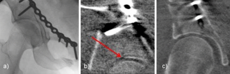
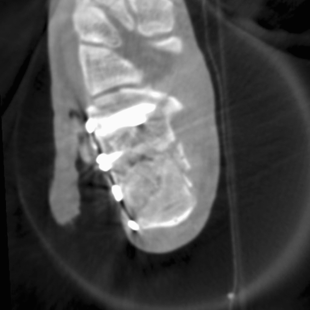
Purpose: Intraoperative 3D imaging has become a valued tool in assessing the quality of reduction and implant placement in orthopedic trauma surgery. In our institution, 3D imaging is used routinely since 2001. To evaluate the intraoperative findings and consequences of this technique, intraoperative revision rates in cases with 3D imaging were analyzed.
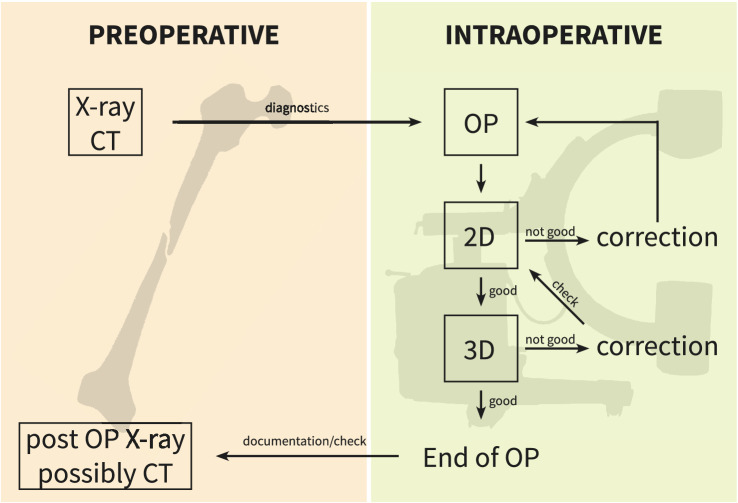
Methods: All operative procedures carried out with intraoperative 3D imaging between August 2001 and December 2016 were included. The scans were assessed intraoperatively and documented thereafter. In case of malreduction or misplaced implants, an immediate revision was performed. The number of scans per case as well as the findings and consequences drawn regarding the anatomical region were analyzed.
Results: 4721 cases with 7201 3D scans were included in this study. The most common anatomical regions were the ankle (22.3%), the calcaneus (14.8%) and the tibial head (9.5%). In 19.1% of all cases, an intraoperative revision was performed. The highest revision rates were found with 36.0% in calcaneal fractures, 24.8% in fractures of the tibial plateau, 22.3% in injuries of the ankle. In 52.0% of revisions, the reduction was improved regarding intra-articular steps or joint congruency. In 30.5% an implant was corrected.
Conclusion: Intraoperative revision due to results of 3D imaging was performed in almost one-fifth of cases. This illustrates the improved possibilities to detect malreduction and implant misplacements intraoperatively and thus the abilities to improve surgical outcome.
Level of evidence: III.
Keywords: 3D imaging; Fractures; Intraoperative imaging; Osteosynthesis.
© 2022. The Author(s).
Conflict of interest statement
The research group, that all authors are part of, receives financial support by Siemens Healthcare (Erlangen, Germany). This did neither influence the concept nor the results of this study.
Figures

Similar articles
-
Intraoperative 3D imaging leads to substantial revision rate in management of tibial plateau fractures in 559 cases.J Orthop Surg Res. 2019 Jul 24;14(1):236. doi: 10.1186/s13018-019-1286-7. J Orthop Surg Res. 2019. PMID: 31340818 Free PMC article.
-
Effect of Intraoperative Three-Dimensional Imaging During the Reduction and Fixation of Displaced Calcaneal Fractures on Articular Congruence and Implant Fixation.Foot Ankle Int. 2015 Jul;36(7):764-73. doi: 10.1177/1071100715576518. Epub 2015 Mar 11. Foot Ankle Int. 2015. PMID: 25761853
-
Intraoperative 3D imaging in the treatment of elbow fractures--a retrospective analysis of indications, intraoperative revision rates, and implications in 36 cases.BMC Med Imaging. 2016 Mar 18;16:24. doi: 10.1186/s12880-016-0126-z. BMC Med Imaging. 2016. PMID: 26987661 Free PMC article.
-
[3D safety in osteosynthesis adjacent to joints].Unfallchirurg. 2016 Oct;119(10):803-10. doi: 10.1007/s00113-016-0228-7. Unfallchirurg. 2016. PMID: 27599821 Review. German.
-
Evolution of imaging in surgical fracture management.Injury. 2020 May;51 Suppl 2:S51-S56. doi: 10.1016/j.injury.2019.10.080. Epub 2019 Oct 24. Injury. 2020. PMID: 31706585 Review.
Cited by
-
Validation of a laser projection platform for the preparation of surgical patches used in paediatric cardiac surgery.Interdiscip Cardiovasc Thorac Surg. 2023 Aug 3;37(2):ivad129. doi: 10.1093/icvts/ivad129. Interdiscip Cardiovasc Thorac Surg. 2023. PMID: 37555820 Free PMC article.
-
Automated Digital Image Optimisation in Intraoperative 2D and 3D Imaging Using a Mobile C-Arm With Flat-Panel Detector.Int J Med Robot. 2025 Apr;21(2):e70053. doi: 10.1002/rcs.70053. Int J Med Robot. 2025. PMID: 40013597 Free PMC article.
References
-
- Vetter SY, Euler F, von Recum J, Wendl K, Gru tzner PA, Franke J. Impact of Intraoperative Cone Beam Computed Tomography on Reduction Quality and Implant Position in Treatment of Tibial Plafond Fractures. Foot & Ankle International [Internet]. 2016 [cited 2017 Mar 2];37:977–82. Available from: http://www.ncbi.nlm.nih.gov/pubmed/27188693 - PubMed
MeSH terms
LinkOut - more resources
Full Text Sources
Medical
Research Materials
