. 2022 Sep 1;17(9):e0273512.
doi: 10.1371/journal.pone.0273512. eCollection 2022.

Multicolor fluorescence activated cell sorting to generate humanized monoclonal antibody binding seven subtypes of BoNT/F

Affiliations

Multicolor fluorescence activated cell sorting to generate humanized monoclonal antibody binding seven subtypes of BoNT/F

Yongfeng Fan et al. PLoS One. .

Abstract

Generating specific monoclonal antibodies (mAbs) that neutralize multiple antigen variants is challenging. Here, we present a strategy to generate mAbs that bind seven subtypes of botulinum neurotoxin serotype F (BoNT/F) that differ from each other in amino acid sequence by up to 36%. Previously, we identified 28H4, a mouse mAb with poor cross-reactivity to BoNT/F1, F3, F4, and F6 and with no detectable binding to BoNT/F2, F5, or F7. Using multicolor labeling of the different BoNT/F subtypes and fluorescence-activated cell sorting (FACS) of yeast displayed single-chain Fv (scFv) mutant libraries, 28H4 was evolved to a humanized mAb hu6F15.4 that bound each of seven BoNT/F subtypes with high affinity (KD 5.81 pM to 659.78 pM). In contrast, using single antigen FACS sorting, affinity was increased to the subtype used for sorting but with a decrease in affinity for other subtypes. None of the mAb variants showed any binding to other BoNT serotypes or to HEK293 or CHO cell lysates by flow cytometry, thus demonstrating stringent BoNT/F specificity. Multicolor FACS-mediated antibody library screening is thus proposed as a general method to generate multi-specific antibodies to protein subtypes such as toxins or species variants.


Conflict of interest statement

The authors have declared that no competing interests exist.

Figures
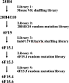
Fig 1
Fig 1. Outline of mAb 28H4 evolution.
mAb 28H4 was evolved to mAb hu6F15.4 with four sequential intermediates (28H4E10, 6F15, hu6F15.1, and hu6F15.3) from five designed single-chain variable fragment (scFv) libraries including two light chain shuffling libraries and three random mutation libraries. MAbs hu6F15.4 and hu6F15.6 showed cross-reactivity to seven BoNT/F subtypes.
Fig 2
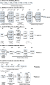
Fig 2. The detailed sorting strategies of each library.
Each library was sorted using FACS at least four rounds using various strategies. Based on the different binding profile of the parental mAbs, libraries were stained with different strategies, including sequentially changed BoNT/F subtype, multiple BoNT/F subtype co-staining (multicolor FACS), or wild type mAb competence (off rate screen). The colonies from last round of sorting were plated on SD-CAA plates for further characterization including DNA sequence analysis and KD and koff measurements. The labels 1st– 9th indicate the round of staining and sorting. Details of staining and sorting conditions are provided in the methods. Library 1. Light chain shuffling of murine scFv 28H4. The library was stained with the indicated individual BoNT/F subtype and sorted for six rounds. This cycle of molecular evolution yielded the scFv 28H4E10. Library 2. Random mutation of scFv 28H4E10. Nine rounds of sorting were performed on the 28H4E10 scFv library. For the first four rounds, aliquots of the library were individually stained with one of the seven BoNT/F HC subtypes and sorted. The individual yeast outputs from sorting were combined and used for the next round of staining and sorting. For the 5th and 6th round of staining and sorting, BoNT/ F1, F2, F5, F6, and F7 HC (Round 5) or BoNT/F1, F2, F5, and F6 HC (Round 6) labeled with unique fluorophores were mixed and used to stain the input yeast. Rounds 7 and 8 were performed as described for Rounds 1–4 and Round 9 was performed after staining with BoNT/F7 HC. This cycle of molecular evolution yielded the scFv 6F15. 3. Library 3. Humanization of scFv 6F15 by light chain shuffling. Five rounds of sorting were performed. For the first round of sorting, the library was stained with a mixture of BoNT/F1 and BoNT/F7 HC labeled with unique fluorophores. For Round 2, the aliquots of the output yeast from Round 1 were individually sorted with one of the indicated BoNT/F HC subtypes and the output from the individual sorting combined. For Rounds 3, 4, and 5, Yeast were stained with a mixture of the indicated BoNT/F HC subtypes labeled with unique fluorophores. This cycle of molecular evolution yielded the scFv hu6F15.1. 4. Library 4. Random mutation of hu6F15.1. Five rounds of sorting were performed. For the first round of staining and sorting, library aliquots were individually stained with either BoNT/F5 HC or a mixture of BoNT/F5 and BoNT/F2 HC and sorted. Sorting outputs were combined and used for the next round of sorting. For Rounds 2–5, yeast were stained with a mixture of the indicated BoNT/F HC subtypes labeled with unique fluorophores. This cycle of molecular evolution yielded the scFv hu6F15.3. 5. Library 5. Random mutation of hu6F15.3. This library was stained and sorted using two approaches. In the first approach, five rounds of sorting were performed. In each round of staining, yeast were stained with a mixture of the indicated BoNT/F HC subtypes labeled with unique fluorophores. A 90-minute period of dissociation of labeled antigen after staining was used during the 3rd and 4th rounds of selection prior to sorting. This cycle of molecular evolution yielded the scFv hu6F15.4. For the second approach, yeast were stained with only BoNT/F7 HC for each round of sorting. A 120-minute period of dissociation of labeled antigen after staining was used during the 4th round of selection prior to sorting. This cycle of molecular evolution yielded the scFv hu6F15.6.
Fig 3
Fig 3. Dot plots of FACS of the mAb 28H4 scFv yeast displayed library stained with a single subtype for each round of sorting.
The 28H4 library was sorted for six rounds sequentially staining with different BoNT subtypes BoNT/F1HC (Rounds 1 and 2) BoNT/F7HC (Round 3) BoNT/F2HC (Round 4), BoNT/F5HC (Round 5), and BoNT/F6HC (Round 6). Each dot represents a single yeast with the BoNT subtype used for staining and the BoNT binding fluorescence shown on the y-axis and the level of scFv yeast surface display on the x-axis. The population gated for sorting and the percent of the total population sorted is shown with the gated yeast colored green.
Fig 4
Fig 4. Dot plots of FACS of the mAb 28H4E10 scFv yeast displayed library using multi-color staining.
A. For the third round of sorting, the scFv library was stained with a mixture of either BoNT subtypes BoNT/F1HC-APCcy-7, BoNT/F5HC-Alexa647, and BoNT/F7HC-Alexa488 or BoNT subtypes BoNT/F1HC-APCcy-7, BoNT/F2HC-PEcy7, and BoNT/F6HC-Pepcy5.5. Each dot represents a single yeast with the BoNT subtype used for staining and the BoNT binding fluorescence shown on the y-axis and the level of scFv yeast surface display on the x-axis. The population gated for sorting and the percent of the total population sorted is shown with the gated yeast colored according to the legend for each of the BoNT subtypes. For sorting the three gates were intersected and only yeast in all three gates (colored blue and with the percent of yeast in the intersected yeast indicated) were collected. B. For the fourth round of sorting, the scFv library was stained with a mixture of BoNT subtypes BoNT/F1HC-APCcy-7, BoNT/F2HC-PEcy7, BoNT/F5HC-Alexa647, BoNT/F6HC-Pepcy5.5, and BoNT/F7HC-Alexa488. Each dot represents a single yeast with the BoNT subtype used for staining and the BoNT binding fluorescence shown on the y-axis and the level of scFv yeast surface display on the x-axis. The population gated for sorting and the percent of the total population sorted is shown with the gated yeast colored according to the legend for each of the BoNT subtypes. For sorting the five gates were intersected and only yeast in all five gates (colored blue and with the percent of yeast in the intersected yeast indicated) were collected.
Fig 5
Fig 5. Specificity of mAbs with broadened specificity and improved affinity.
A. Yeast displaying the indicated scFv were stained with either an equimolar mixture of BoNT serotypes A, B, C, D, E, and G or with BoNT/F1. Each yeast displayed scFv bound BoNT/F1 but did not bind the mixture of the other six serotypes. B. Yeast displaying the indicated scFv with stained with either biotinylated HEK293 or Chinese Hamster Ovary (CHO) cell lines or with biotinylated BoNT/F2HC as a positive control. Binding was detected using streptavidin-PE. scFv did not bind HEK or CHO cell lines but did bind BoNT/F2 in proportion to their affinity.
Fig 6
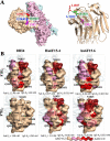
Fig 6. The epitopes of 28H4, hu6F15.4 and hu6F15.6 on BoNT/F1 and BoNT/F7.
A. The key amino acid residues in the epitope of 28H4, hu6F15.4 and hu6F15.6. In the left panel, four key residues for the binding of 28H4 to BoNT/F1 (T993, R1026, L1027 and G1048) are shown on a surface model of the BoNT/F1 holotoxin (Alphafold, AF-A7GBG3). The BoNT/F1 HC is shown in beige, the catalytic domain (LC) in blue, and the translocation domain in pink. In the right panel, the side chains of the same four key residues are shown on a ribbon diagram of the crystal structure of BoNT/F1 HC (pdb, 3fuq). The four key amino acid residues are located on three different loops B. The fine epitope maps of mAbs 28H4. The contribution of amino acid side chains in mAb binding was measured and shown by coloring the surface side chain projection on either the BoNT/F1HC structure (pdb, 3fuq) or a model of the BoNT/F7HC based on 3fuq. Surface side chains were colored based on the values of ΔΔG from beige (no impact on mAb binding, ΔΔG<0.2) to dark red, ΔΔG>2.0 according to the key shown at the bottom of the Figure.

References

    1. Garcia-Rodriguez C, Geren I, Lou J, Conrad F, Forsyth C, Wen W, et al.. Neutralizing human monoclonal antibodies binding multiple serotypes of botulinum neurotoxin. Protein Engineering, Design & Selection. 2011;24(3):321–31. doi: 10.1093/protein/gzq111 - DOI - PMC - PubMed
    1. Meyer RF, Miller L, Bennett RW, MacMillan JD. Development of a monoclonal antibody capable of interacting with five serotypes of Staphylococcus aureus enterotoxin. Applied Enviro Micro. 1984;47(2):283–7. doi: 10.1128/aem.47.2.283-287.1984 - DOI - PMC - PubMed
    1. Flyak AI, Shen X, Murin CD, Turner HL, David JA, Fusco ML, et al.. Cross-Reactive and Potent Neutralizing Antibody Responses in Human Survivors of Natural Ebolavirus Infection. Cell. 2016;164(3):392–405. doi: 10.1016/j.cell.2015.12.022 - DOI - PMC - PubMed
    1. Rockx B, Corti D, Donaldson E, Sheahan T, Stadler K, Lanzavecchia A, et al.. Structural Basis for Potent Cross-Neutralizing Human Monoclonal Antibody Protection against Lethal Human and Zoonotic Severe Acute Respiratory Syndrome Coronavirus Challenge. J Virol. 2008;82(7):3220–35. doi: 10.1128/JVI.02377-07 - DOI - PMC - PubMed
    1. Simmons CP, Bernasconi NL, Suguitan AL Jr, Mills K, Ward JM, Chau NVV, et al.. Prophylactic and therapeutic efficacy of human monoclonal antibodies against H5N1 influenza. PLoS Med. 2007;4(5). doi: 10.1371/journal.pmed.0040178 - DOI - PMC - PubMed

Publication types