. 2022 Sep 2;377(6610):eabq4515.
doi: 10.1126/science.abq4515. Epub 2022 Sep 2.

GnRH replacement rescues cognition in Down syndrome

Affiliations

GnRH replacement rescues cognition in Down syndrome

Maria Manfredi-Lozano et al. Science. .

Abstract

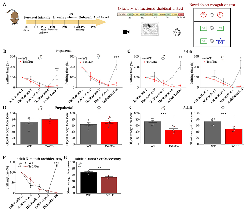
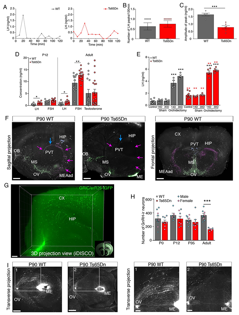
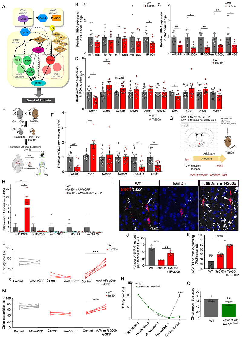
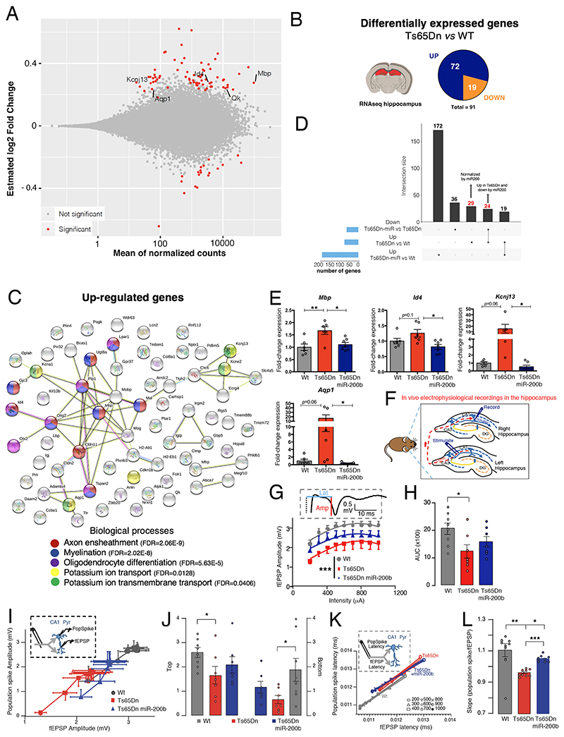
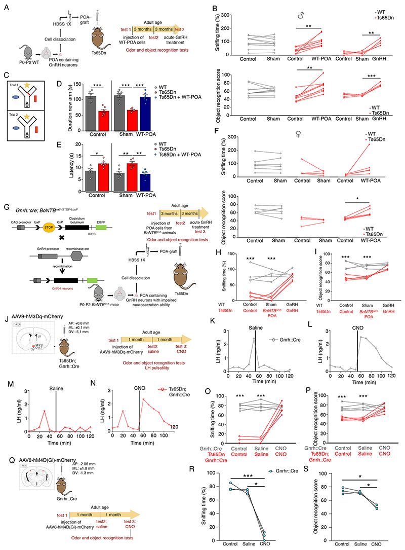
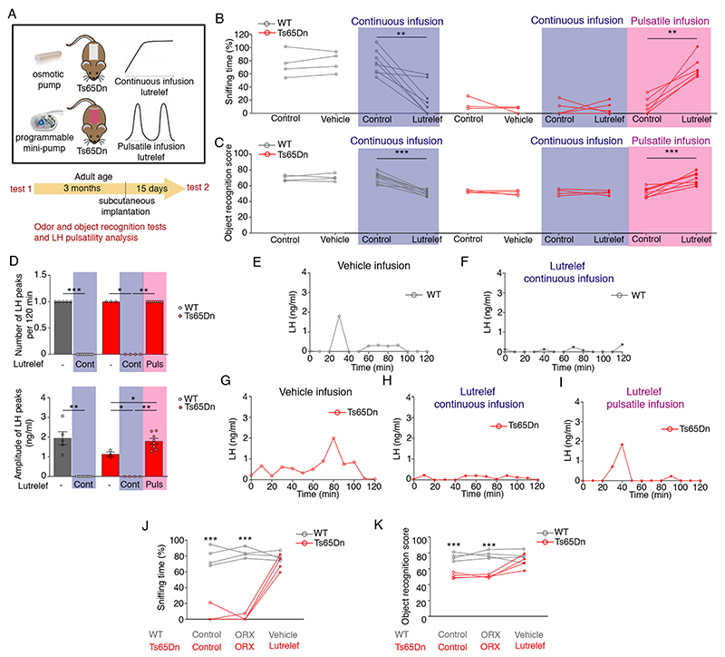
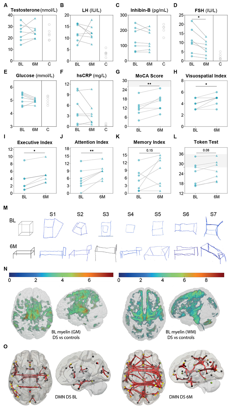
At the present time, no viable treatment exists for cognitive and olfactory deficits in Down syndrome (DS). We show in a DS model (Ts65Dn mice) that these progressive nonreproductive neurological symptoms closely parallel a postpubertal decrease in hypothalamic as well as extrahypothalamic expression of a master molecule that controls reproduction-gonadotropin-releasing hormone (GnRH)-and appear related to an imbalance in a microRNA-gene network known to regulate GnRH neuron maturation together with altered hippocampal synaptic transmission. Epigenetic, cellular, chemogenetic, and pharmacological interventions that restore physiological GnRH levels abolish olfactory and cognitive defects in Ts65Dn mice, whereas pulsatile GnRH therapy improves cognition and brain connectivity in adult DS patients. GnRH thus plays a crucial role in olfaction and cognition, and pulsatile GnRH therapy holds promise to improve cognitive deficits in DS.


Conflict of interest statement

Competing interests: M.M.-L., V.L., A.M. P.G. and V.P. disclose that they are inventors of a patent covering the treatment of cognitive disorder and dementia with pulsatile GnRH (82). All other authors do not have competing interests.

Figures

Fig. 1
Fig. 1. Ts65Dn mice show age-dependent olfactory, cognitive loss.
(A) Experimental design to evaluate olfactory and visual discrimination during postnatal development. (B-E) Habituation/dishabituation test to assess the ability of P35 and adult Ts65Dn mice and WT littermates to differentiate between two distinct odors (P35: B: WT male dishabituation vs. Ts65Dn male dishabituation, t(45) = 3.67, P = 0.003, n=6,5; WT female dishabituation vs. Ts65Dn female dishabituation, t(105) = 6.10, P < 0.0001, n=6,5. Adult: C: WT male dishabituation vs. Ts65Dn male dishabituation, t(45) = 4.19, P = 0.0006, n=6,5; WT female dishabituation vs. Ts65Dn female dishabituation, t(16) = 5.42, P = 0.0002, n=5,5); or recognize new objects in their environment (P35: D: WT male vs. Ts65Dn male, t(9) = 1.02, P = 0.33, n=6,5; WT female vs. Ts65Dn female, t(16) = 1.278, P = 0.22, n=9,9. E: WT male vs. Ts65Dn male, t(10) = 4.8, P = 0.0007, n=6,6; WT female vs. Ts65Dn female, t(10) = 4.93, P = 0.0006, n=6,6). Values represent means ± SEM. Unpaired Student’s t-test to compare 2 conditions; two-way repeated-measures ANOVA followed by Sidak’s post hoc test for ≥ 3 conditions. **P<0.01; ***P<0.001.
Fig. 2
Fig. 2. Ts65Dn mice progressively lose GnRH expression, function.
(A) LH pulsatility assessment by serial blood sampling. (B) LH pulse frequency (n=8,8 mice). (C) LH pulse amplitude (n=5,6 mice). (D) Circulating levels of LH, FSH (at P12 and in adults) and testosterone (in adults) (WT n=8,12,8,13,15 mice; Ts65Dn n=5,12,9,14,20 mice). (E) Effect of orchidectomy on LH levels 14 days (14D) and 30 days (30D) after surgery (WT: n=8,4,4,4,4 mice; Ts65Dn: n=9,4,4,5,5 mice). (F) GnRH-immunoreactive neuronal fiber tracing, pseudocolored projection pathways. Green: GnRH neurons and projections to median eminence (ME); magenta: neuronal projections to the anterodorsal amygdala (MEAad); blue: neuronal projections to the paraventricular thalamus (PVT). CX: cortex; HIP: hippocampus; MS: medial septum; OB: olfactory bulb; OV: organum vasculosum laminae terminalis; PVT: paraventricular thalamus. Scale bars: sagittal 350 μm; frontal 500 μm. (G) Three-dimensional imaging of solvent-cleared organs (iDISCO) showing Cre-dependent Tau-GFP expression in mouse neurons of the CX and HIP, under the control of the GnRH receptor (Gnrhr) promoter. Scale bar: 200 μm (H) GnRH-immunoreactive cell bodies in the preoptic region of WT and Ts65Dn mice at different postnatal ages assessed by conventional neuroanatomical analyses (WT, n=9,6,8,8 mice; Ts65Dn n=8,8,7,6 mice). (I) Representative horizontal view of whole-mount GnRH immunoreactivity in adult (P90) Ts65Dn and WT littermates followed by iDISCO. Scale bars: 600 μm (panels 1, 2: 300 μm). Values represent means ± SEM. *P<0.05; **P< 0.01; ***P<0.001. Unpaired Student’s t-test (C,D,Ered-asterisks,H) or Mann-Whitney U test for comparison between 2 conditions (B,D); one-way ANOVA followed by Tukey’s post hoc test for ≥ 3 conditions (E). Red asterisks indicate a comparison between genotypes.
Fig. 3
Fig. 3. GnRH transcriptional machinery disequilibrium underlies cognitive impairments.
(A) Putative microRNA-transcription-factor network regulating hypothalamic GnRH promoter activation during postnatal development (adapted from (18)). (B-D) RT-PCR analysis of expression levels of microRNAs located on chromosome 16 (B) and miR-200 family members (C), as well as hypothalamic GnRH promoter modulators (D) in the preoptic region (POA) of adult WT and Ts65Dn littermates (B: n=11,10,10,11,10 WT mice; n=9,8,8,9,8 Ts65Dn mice; C: n=8,9,7,9,9 WT mice; n=6,6,6,6,6 Ts65Dn mice; D: n=11,11,9,10,11,11,7,11,11,8 WT mice; n=9,9,9,9,9,9,6,9,9,6 Ts65Dn mice). (E) Generation of Gnrh::Gfp;Ts65Dn reporter mice, which express GFP under an ectopic Gnrh promoter. GnRH-GFP neurons were FACS-isolated from the POA of Gnrh::Gfp and Gnrh::Gfp;Ts65Dn littermates at P12. (F) RT-PCR analysis of gene expression in FACS-sorted GnRH-GFP cells (n=10,10,11,11,10,11 WT mice; n=9,7,9,9,9,10 Ts65Dn mice). (G) Experimental design to evaluate the functional involvement of miR-200 family members in odor discrimination and novel object recognition in Ts65Dn mice. Red dot: viral injection site; LV, lateral ventricle; MePO, median preoptic nucleus; OVLT, organum vasculosum laminae terminalis. (H) Effect of viral overexpression of miR-200b in the POA on miR-200 family member expression (n=4,5,5 mice). (I-M) Effect of viral miR-200b overexpression in the POA on the number of neurons expressing Gnrh transcripts in the OVLT (I,J) and the proportion expressing Otx2 (I,K), as assessed by fluorescent in situ hybridization, as well as odor discrimination (L) and novel object recognition (M) in Ts65Dn mice (J,K; n=4,4,4 mice; L,M: n=5,5,6 mice). (N,O) Odor discrimination (N; n=6 per group) and novel object recognition (O; n=6 per group) in 12-month old male mice selectively lacking Dicer in GnRH neurons. Values represent means ± SEM. *P<0.05; **P<0.01; ***P<0.001. Unpaired Student’s-test or Mann-Whitney U test (B-D,F), paired Student’s-test or Wilcoxon matched-pair test (L,M) and Kruskal-Wallis test (H) for comparisons between 2 conditions, one-way (J,K) or two-way repeated measures (N,O) ANOVA followed by Tukey’s and Sidak’s post hoc tests for ≥ 3 conditions.
Fig. 4
Fig. 4. Hypothalamic miR-200b overexpression rescues hippocampal transcriptome, connectivity.
(A)MA plot of gene expression changes (estimated log2 fold changes as a function of the mean of normalized counts; PAdj<0.05) in the hippocampus of adult male (P180) Ts65Dn (n=3) vs. WT mice (n=4). (B) Pie chart of the number of differentially regulated genes between Ts65Dn and WT littermates in the hippocampus (Padj<0.05). (C) STRING protein network analysis of upregulated genes. (D) UpSet plot showing the intersection between differentially up-regulated genes in the Ts65Dn hippocampus and genes rescued by miR-200b. (E) Quantitative RT-PCR confirmation of RNA-seq data. (F) Schematic diagram illustrating in vivo electrophysiological recordings in the dorsal hippocampus of adult WT (n=8), Ts65Dn (n=7) and Ts65Dn mice with miR-200b overexpression (n=7). Field excitatory postsynaptic potentials (fEPSPs) and population spikes were evoked in the hippocampal CA1 area by stimulating commissural fibers in the contralateral hippocampus. (G) Synaptic input-output (I/O) curves indicating the relationship between fEPSP amplitude at increasing stimulus intensities (200-1000 μA). Insert: Representative fEPSP recording with measurement of latency (blue line) and amplitude (red line). (H) Area under the curve (AUC) of fEPSP responses. (I) Boltzmann-fitted fEPSP-population spike coupling. Insert: schematic showing both fEPSP and population spike recording in the CA1 pyramidal layer (CA1 pyr) following the same commissural path stimulation. (J) Top and bottom Boltzmann-fitted parameters of fEPSP-population spike coupling as a measure of intrinsic excitability of CA1 pyramidal neurons. (K) Relationship between the mean fEPSP latency and population spike latency at different stimulus intensities (200-1000 μA). Insert: schematic showing both fEPSP and population spike recording in the CA1 pyramidal layer (CA1 pyr) following the same commissural path stimulation. (L): Slopes of the fEPSP-population spike relationships. Data represent means ± SEM. *P ≤ 0.05; **P ≤ 0.01;***P≤0.001. Kruskal-Wallis ANOVA and Mann-Whitney U test (G) for comparisons between two conditions, one-way ANOVA followed by Tukey’s post hoc test (E,H,L) or two-way ANOVA (J) for ≥ 3 conditions.
Fig. 5
Fig. 5. Restoring GnRH neurons/function reverses olfactory, cognitive deficits.
(A) Cell therapy by grafting enzymatically dissociated cells from the POA of WT neonatal mice (P0-P2) into the third ventricle of adult Ts65Dn mice. (B-F) Effect of WT-POA grafts in Ts65Dn males on olfactory and cognitive performance (B: n=10,9,5 male mice; F: n=7,5,5 female mice) and short-term visuospatial memory assessed by the Y-maze test (C-E) 3 months after surgery (D: n=5,6,7,5,7 male mice; E: n=5,6,7,5,6 male mice). (G) Experimental design to graft POA cells from neonatal mice with exocytotis-incompetent GnRH neurons (Gnrh::Cre; BoNTBloxP-STOP-loxP). (H,I) After a 3-month recovery period, effect of BoNTBGnrh-POA grafts and acute intraperitoneal GnRH injection on odor discrimination (H) and object recognition (I) (H,I: n=4,5 mice). (J) Experimental design to study LH pulsatility, cognitive and olfactory performance after the chemogenetic activation of GnRH neurons by injecting adult Gnrh::Cre and Ts65Dn;Gnrh::Cre mice with an hM3Dq DREADD viral vector followed by CNO (clozapine N-oxide solution; 1mg/kg of body weight) (n=5,5 mice). Red dots: virus injection sites. 3V: third ventricle; LV: lateral ventricle; MePO: median preoptic nucleus; OVLT: organum vasculosum laminae terminalis. (K-N) Representative graphs for LH pulsatility; (O) odor discrimination; (P) novel object recognition. (Q) Experimental design to study olfactory and cognitive performance before and after the chemogenetic inhibition of GnRH-R expressing neurons in 6-month old Gnrhr::Cre mice by injection of an hM4D(Gi) DREADD viral vector. Red dots: virus injection sites. (R) Odor discrimination; (S) novel object recognition (n=3,3 mice). Values represent means ± SEM. * P<0.05,** P<0. 01,***P<0.001; paired Student’s t-test or Wilcoxon matched-pair test (F); one-way ANOVA (D,E) or oneway (R,S) or two-way (B,H,I,O,P) repeated-measures ANOVA followed by Tukey’s or Sidak’s post hoc tests, respectively.
Fig. 6
Fig. 6. Olfactory, cognitive deficit reversal requires GnRH pulsatility.
(A) Schematic of pharmacotherapy with Lutrelef, a clinically-used GnRH peptide, in adult Ts65Dn mice. Mice were implanted with osmotic pumps to receive a continuous infusion of vehicle or Lutrelef (0.25 μg/3h over 2 weeks), or with a programmable mini-pump (iPRECIO), to receive pulsatile Lutrelef infusion (0.25 μg/10 min every 3 hours over 2 weeks). (B-I) Effect of treatments on odor discrimination (B; n=4,8,3,5,6 mice), object recognition (C; n=4,9,4,4,8 mice) and LH pulsatility (D-I) (D: n=4,9,3,4,8 mice; each dot represents one subject). (J-K) Effects of Lutrelef in orchidectomized (ORX) mice (n=4). Values represent means ± SEM. * P<0.05,** P<0. 01,***P<0.001; paired Student’s t-test or Wilcoxon matched-pair test (B,C), unpaired Student’s t-test or Mann-Whitney U-test (D) or a two-way repeated-measures ANOVA followed by Sidak’s post hoc test (J,K).
Fig. 7
Fig. 7. Pulsatile GnRH improves patient brain connectivity, cognition.
(A-F) Biochemical profile at baseline (BL, blue dots, n=7) and after 6-month pulsatile GnRH therapy (6M, blue triangles, n=7), compared to healthy age- and sex-matched controls (C, open circles, n=5 for inhibin-B and n=6 for all the other parameters). (G-L) Results of cognitive tests in male Down syndrome patients (n=7) at BL (blue dots) and after 6M of GnRH therapy (blue triangles): MoCA total score, visuo-spatial index, executive index, attention index, memory index and Token test score. (M) 3D-drawings representing a cube and a bed, part of visuospatial index score of the MoCA at BL and 6M for each subject (S1-S7). S2, S3, S5 and S7 improved at 6M. (N) Statistical parametric maps of brain anatomy differences between DS patients (n=8) and age-matched male controls (n=44) after pFWE<0.05 correction for multiple comparisons at the whole-brain level. Voxel-based quantification of magnetization transfer (MT) saturation maps reveal volume loss in the cerebellum, anterior cingulate cortex, supplementary motor cortex, substantia nigra, thalamus, insula and primary motor cortex M1, and loss of myelin content in the thalamus, primary sensorimotor cortex S1/M1, angular gyrus, insula, and superior frontal and temporal gyri bilaterally. (O) Resting-state functional MRI comparison of functional connectivity in DS (n=7) at BL and after 6MGnRH therapy in the visual (including all occipital regions, the lingual gyrus and the cuneus) and sensorimotor (pre- and post-central gyri, middle frontal gyrus) default-mode network (DMN), connected to the superior parietal lobule, the superior temporal gyrus, some prefrontal areas and part of the anterior DMN (increased; FDR-corrected p<0.0005); as well as within the hippocampal regions of the ventral DMN linked to the amygdala (reduced; FDR-corrected p<0.0005). *p<0.05, **p<0.01. MoCA: Montreal cognitive assessment; FDR: false discovery rate; FWE: family-wise error; GM: grey matter; WM: white matter.

Comment in

References

    1. Antonarakis SE, et al. Down syndrome. Nat Rev Dis Primers. 2020;6:9. - PMC - PubMed
    1. de la Torre R, et al. Safety and efficacy of cognitive training plus epigallocatechin-3-gallate in young adults with Down’s syndrome (TESDAD): a double-blind, randomised, placebo-controlled, phase 2 trial. Lancet Neurol. 2016;15:801–810. - PubMed
    1. Shapiro BL. Down syndrome--a disruption of homeostasis. Am J Med Genet. 1983;14:241–269. - PubMed
    1. Baburamani AA, Patkee PA, Arichi T, Rutherford MA. New approaches to studying early brain development in Down syndrome. Dev Med Child Neurol. 2019;61:867–879. - PMC - PubMed
    1. Bull MJ. Down Syndrome. N Engl J Med. 2020;382:2344–2352. - PubMed

MeSH terms

Substances