. 2022 Jun 21;3(3):970-974.
doi: 10.1002/jha2.479. eCollection 2022 Aug.

CAR-T cells derived from multiple myeloma patients at diagnosis have improved cytotoxic functions compared to those produced at relapse or following daratumumab treatment

Affiliations

CAR-T cells derived from multiple myeloma patients at diagnosis have improved cytotoxic functions compared to those produced at relapse or following daratumumab treatment

Audrey Abecassis et al. EJHaem. .

Abstract

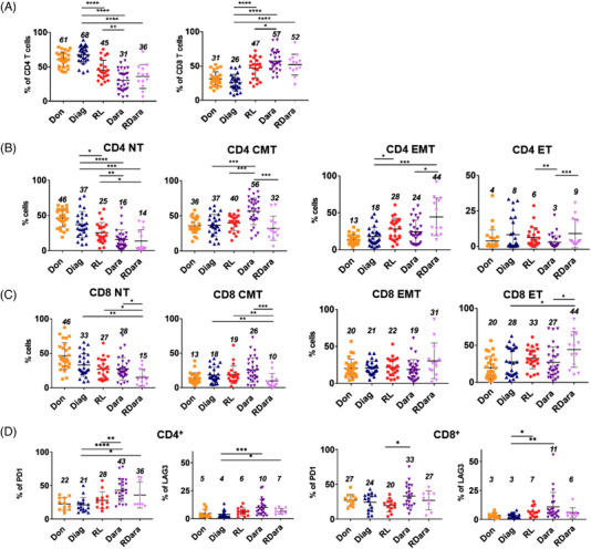
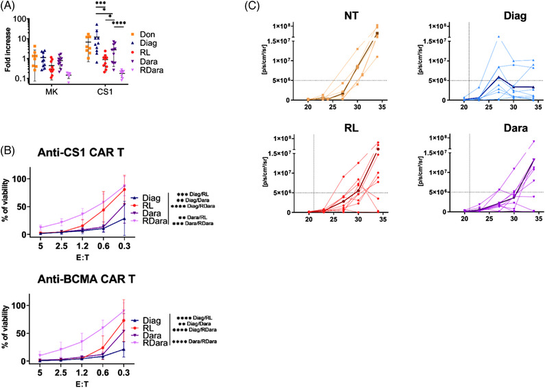
Chimeric antigen receptor T cells (CAR-T) have provided promising results in multiple myeloma (MM). However, many patients still relapse, pointing toward the need of improving this therapy. Here, we analyzed peripheral blood T cells from MM patients at different stages of the disease and investigated their phenotype and capacity to generate functional CAR-T directed against CS1 or B Cell Maturation antigen. We found a decrease in naive T cells and elevated frequencies of exhaustion markers in T cells from treated MM patients. Interestingly, individuals treated with daratumumab display elevated ratios of central memory T cells. CAR-T derived from patients at relapse show reduced in vitro expansion and cytotoxic capacities in response to MM cells compared to those produced at diagnosis. Of note, CAR-T from daratumumab treated patients display intermediate defects. Reduced anti-myeloma activity of CAR T cells from treated patients was also observed in a mouse model. Our findings suggest that T cell defects in MM patients, specifically during relapse, have a major impact on their capacity to generate efficient therapeutic CAR-T. Selecting naive or central memory T cell subsets to generate therapeutic T cells could improve the CAR-T therapy for MM.

Keywords: CAR‐T cells; daratumumab; multiple myeloma; peripheral tcells; relapse.


Conflict of interest statement

The authors declare no competing financial interests and have no conflict of interest to disclose.

Figures

FIGURE 1
FIGURE 1
Analysis of T‐cell subsets in the peripheral blood of multiple myeloma (MM) patients. (A) Scatter dot plots show the percentages of CD4 and CD8 expressing T cells in peripheral blood mononuclear cells (PBMCs) isolated from healthy donors (Don, N = 27), MM patients at diagnosis (Diag, N = 26), at relapse MM (RL, N = 23), treated with daratumumab (Dara, N = 30), and in relapse after daratumumab treatment (RDara, N = 13). (B and C) Scatter dot plots show the percentages of naïve (CD3+, CD62L+, CD45RO‐) (N), effector (CD3+, CD62L‐, CD45RO‐) (E), central memory (CD3+, CD62L+, CD45RO+) (CM) and effector memory (CD3+, CD62L‐, CD45RO+) (EM) among CD4 (B) and CD8 (C) expressing T cells from healthy donors and MM patients as in (A). (D) Scatter dot plots show the percentages of PD1 and LAG3 expressing cells among CD4 or CD8 positive CD3+ cells in Don (N = 12), Diag (N = 16), RL (N = 13), Dara (N = 23), and RDara (N = 9). Numbers in the scatter dot plots indicate the mean percentages of each cohort. p‐values between two groups were examined with a Mann‐Whitney U‐test. *p ≤ 0.05, **p ≤ 0.005, ***p ≤ 0.001
FIGURE 2
FIGURE 2
Functional analysis of chimeric antigen receptor T cells (CAR‐T) from multiple myeloma (MM) patients. (A) Scatter dot plot showing expansion (total number/starting number) of CAR‐T cells cultured for 7 days on NIH3T3 cells expressing the CS1 antigen. Anti‐CS1 CAR‐T (CS1) or nonfunctional CAR‐T (MK) generated from donors (Don, N = 8) and MM patients at diagnosis (Diag, N = 10), in relapse (RL, N = 10), treated with dartumumab (Dara, N = 11) or in relapse after daratumumab treatment (RDara, N = 8) were expanded with anti‐CD3/anti‐CD28 beads and interleukine‐2 (IL‐2) for 6 days, then separated from beads/IL‐2, and 5.104 cells were plated on feeders. (B) In vitro anti‐MM activity of anti‐CS1 and anti‐BCMA CAR‐T cells from MM patients. Curves represent the percentage of viable MM1.S cells in the culture compared to controls with no specific killing. 2.5 × 104 luciferase expressing MM1.S cells (Target:T) were co‐cultured with anti‐CS1 CAR‐T (Effector: E) from MM patients, at various E:T ratios for 24 h. Anti‐CS1 CAR‐T were generated from patients at diagnosis (Diag, N = 8), at relapse (RL, N = 8), upon daratumumab (Dara, N = 9) or in relapse after daratumumab treatment (RDara, N = 8). Anti‐B cell maturation antigen (BCMA) CAR‐T were generated from patients at diagnosis (Diag, N = 4), at relapse (RL, N = 4), upon daratumumab (Dara, N = 6) or in relapse after daratumumab treatment (RDara, N = 8). p‐values were examined with a two‐way ANOVA test (*p < 0.05; **p < 0.01; ***p < 0.001; ****p < 0.0005). (C) In vivo activity of anti‐CS1 CAR‐T generated from MM patients in the MM1.Sluc Xenograft mouse model. Shown are the longitudinal radiance levels (luciferase activity) measured in mice injected with CAR‐T (dotted line) from MM patients (Diag, RL, Dara) or left untreated (NT). Six to 12‐week‐old NOD/SCID/IL‐2Rγnull mice were inoculated with 5 × 106 MM1.SLuc cells by tail vein injection at day 0, followed, 21 days later, by infusion of 106 CAR‐T. Bioluminescence was measured with the IVIS Imaging System at day 20, 23, 26, 30, and 34 after tumor injection. Bold curves represent the average radiances for each cohort. Radiance was measured on the entire body of mice. These experiments were performed with CAR‐T generated from three independent patients of each group, each injected into three independent mice (nine mice per group)

References

    1. Gay F, Engelhardt M, Terpos E, Wäsch R, Giaccone L, Auner HW, et al. From transplant to novel cellular therapies in multiple myeloma: European Myeloma Network guidelines and future perspectives. Haematologica. 2018;103:197–211. 10.3324/haematol.2017.174573 - DOI - PMC - PubMed
    1. Dimopoulos MA, Oriol A, Nahi H, San‐Miguel J, Bahlis NJ, Usmani SZ, et al. Daratumumab, lenalidomide, and dexamethasone for multiple myeloma. N Engl J Med. 2016;375:1319–31. 10.1056/NEJMoa1607751 - DOI - PubMed
    1. Facon T, Kumar S, Plesner T, Orlowski RZ, Moreau P, Bahlis N, et al. Daratumumab plus lenalidomide and dexamethasone for untreated myeloma. N Engl J Med. 2019;380:2104–15. 10.1056/NEJMoa1817249 - DOI - PMC - PubMed
    1. Carpenter RO, Evbuomwan MO, Pittaluga S, Rose JJ, Raffeld M, Yang S, et al. B‐cell maturation antigen is a promising target for adoptive T‐cell therapy of multiple myeloma. Clin Cancer Res. 2013;19:2048–60. 10.1158/1078-0432.CCR-12-2422 - DOI - PMC - PubMed
    1. Chu J, He S, Deng Y, Zhang J, Peng Y, Hughes T, et al. Genetic modification of T cells redirected toward CS1 enhances eradication of myeloma cells. Clin Cancer Res. 2014;20:3989–4000. 10.1158/1078-0432.CCR-13-2510 - DOI - PMC - PubMed