

Solitary fibrous tumor of the liver: A case report and review of the literature
- PMID: 36051139
- PMCID: PMC9297403
- DOI: 10.12998/wjcc.v10.i20.7097
Solitary fibrous tumor of the liver: A case report and review of the literature
Abstract
Background: Hepatic solitary fibrous tumor (SFT) is a rare neoplasm. Up to now, only 90 cases have been reported in the English language literature. This report describes a case of SFT of the liver misdiagnosed as hepatocellular carcinoma.
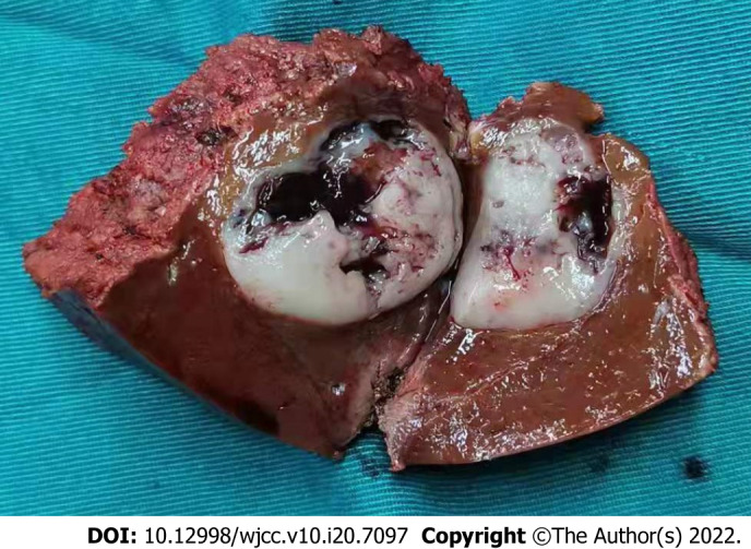
Case summary: A 42-year-old male had a two-year history of a gradually enlarging intrahepatic nodule. The preoperative imaging revealed a mass with a size of 2.7 cm × 2.3 cm located in the segment IV of the liver. The patient was subjected to the resection of the segment IV, such as the medial segment of the left lobe of the liver. The histological examination of the mass showed various spindled cells irregularly arranged in the stroma. The immunohistochemistry of this mass revealed a positive staining for CD34 and STAT6. The history of intracranial tumor and postoperative pathological results led to the diagnosis of SFT of the liver (SFTL) due to a metastasis from the brain.
Conclusion: SFTL is an uncommon mesenchymal neoplasm that can be easily overlooked or misdiagnosed. The best treatment choice is the complete surgical resection of the mass. A regular follow-up after the surgery should be performed due to the poor prognosis of metastatic or recurrent SFT.
Keywords: Case report; Liver; Mesenchymal neoplasm; Metastasis; Solitary fibrous tumor; Surgical treatment.
©The Author(s) 2022. Published by Baishideng Publishing Group Inc. All rights reserved.
Conflict of interest statement
Conflict-of-interest statement: The authors declare that they have no conflict of interest.
Figures


References
-
- Klemperer P, Coleman BR. Primary neoplasms of the pleura. A report of five cases. Am J Ind Med. 1992;22:1–31. - PubMed
-
- Louis DN, Perry A, Reifenberger G, von Deimling A, Figarella-Branger D, Cavenee WK, Ohgaki H, Wiestler OD, Kleihues P, Ellison DW. The 2016 World Health Organization Classification of Tumors of the Central Nervous System: a summary. Acta Neuropathol. 2016;131:803–820. - PubMed
-
- Soussan M, Felden A, Cyrta J, Morère JF, Douard R, Wind P. Case 198: solitary fibrous tumor of the liver. Radiology. 2013;269:304–308. - PubMed
-
- Demicco EG, Wagner MJ, Maki RG, Gupta V, Iofin I, Lazar AJ, Wang WL. Risk assessment in solitary fibrous tumors: validation and refinement of a risk stratification model. Mod Pathol. 2017;30:1433–1442. - PubMed
Publication types
LinkOut - more resources
Full Text Sources
Research Materials
Miscellaneous
