Pseudoshock: A Challenging Presentation of Bilateral Subclavian Artery Stenosis
- PMID: 36051171
- PMCID: PMC9426959
- DOI: 10.12890/2022_003495
Pseudoshock: A Challenging Presentation of Bilateral Subclavian Artery Stenosis
Abstract
Introduction: Subclavian artery stenosis (SAS) is a manifestation of peripheral artery disease (PAD). Presentation varies, ranging from arm claudication and muscle fatigue to symptoms which reflect vertebrobasilar hypoperfusion, among which are syncope, ataxia and dysphagia. Although rare, severe bilateral SAS can exist and present as refractory hypotension. We describe a case of bilateral SAS masquerading as circulatory shock, or rather 'pseudoshock'.
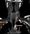
Case description: A 59-year-old female patient presented to the emergency department with complaints of dark stools. She was anaemic and hypotensive and therefore suspected to have an acute gastrointestinal bleed (GIB) with resultant haemorrhagic shock. Her hypotension was unresponsive to fluid resuscitation and blood transfusions. Bilateral upper extremity radial artery catheters confirmed low blood pressures. After her blood pressure failed to improve despite the addition of several vasopressors, a femoral artery catheter (FAC) was placed, which revealed significant hypertension discordant with the hypotension measured by the radial artery catheters. Review of CT angiography of the upper extremities revealed the presence of bilateral SAS which was deemed to be the aetiology of the falsely low blood pressure.
Discussion: SAS should be suspected in patients with lower extremity PAD or a blood pressure (BP) differential of 15 mmHg or more between arms. When bilateral subclavian arteries are stenosed, this difference in BP may be concealed, making lower extremity BP measurements, as seen in non-invasive tests such as ankle brachial index (ABI) tests or through more invasive procedures such as FAC placement, critically important.
Conclusion: Bilateral SAS may present as pseudo-hypotension. In cases of refractory shock of unclear aetiology, especially in patients with known PAD, a high index of suspicion is warranted for 'pseudoshock' secondary to severe vascular stenosis. Comparison of upper and lower extremity BP via invasive arterial catheters or non-invasive ABI tests can aid in the diagnosis of bilateral SAS.
Learning points: Bilateral subclavian artery stenosis (SAS) may present as pseudo-hypotension and shock of unclear aetiology.In patients with underlying peripheral arterial disease, pseudoshock should be considered in the differential diagnosis.Comparison of upper and lower extremity blood pressure via invasive arterial catheters or the non-invasive ankle brachial index (ABI) test has diagnostic value for bilateral SAS.Pseudoshock is managed via secondary prevention with antiplatelets and statins for asymptomatic patients, and revascularization for symptomatic patients.
Keywords: Bilateral subclavian artery stenosis; peripheral vascular disease; pseudoshock; shock; vasopressors; vertebrobasilar insufficiency.
© EFIM 2022.
Conflict of interest statement
Conflicts of Interests: The authors declare there are no competing interests.
Figures
References
-
- Ramin S, Michael HC, Warner PB, Arnost F, Julie OD, et al. Subclavian artery stenosis: prevalence, risk factors, and association with cardiovascular diseases. J Am Coll Cardiol. 2004;44:618–623. - PubMed
-
- Hirata K, Nakazato J, Wake M, Takahashi T. Pseudo-hypotension with acute pulmonary oedema due to simultaneous bilateral subclavian artery stenosis in a patient with coronary artery bypass graft surgery using bilateral internal mammary arteries: a case report. Oxf Med Case Reports. 2019;2019:omz038-omz. - PMC - PubMed
-
- Caesar-Peterson S, Bishop MA, Qaja E. Subclavian artery stenosis. Treasure Island (FL): StatPearls Publishing; 2021. - PubMed
-
- Standring S, Gray H, Borley N. Gray’s anatomy: the anatomical basis of clinical practice. 40th ed. Amsterdam: Elsevier; 2021.
-
- Rahimi O, Geiger Z. Anatomy, thorax, subclavian arteries. Treasure Island (FL): StatPearls Publishing; 2021. - PubMed
LinkOut - more resources
Full Text Sources


