. 2022 Aug 16:13:990993.
doi: 10.3389/fphar.2022.990993. eCollection 2022.

Protective effect of quercetin on cadmium-induced renal apoptosis through cyt-c/caspase-9/caspase-3 signaling pathway

Affiliations

Protective effect of quercetin on cadmium-induced renal apoptosis through cyt-c/caspase-9/caspase-3 signaling pathway

Ruxue Huang et al. Front Pharmacol. .

Abstract

Cadmium (Cd), a heavy metal, has harmful effects on animal and human health, and it can also obviously induce cell apoptosis. Quercetin (Que) is a flavonoid compound with antioxidant and other biological activities. To investigate the protective effect of Que on Cd-induced renal apoptosis in rats. 24 male SD rats were randomly divided into four groups. They were treated as follows: control group was administered orally with normal saline (10 ml/kg); Cd group was injected with 2 mg/kg CdCl2 intraperitoneally; Cd + Que group was injected with 2 mg/kg CdCl2 and intragastric administration of Que (100 mg/kg); Que group was administered orally with Que (100 mg/kg). The experimental results showed that the body weight of Cd-exposed rats significantly decreased and the kidney coefficient increased. In addition, Cd significantly increased the contents of Blood Urea Nitrogen, Creatinine and Uric acid. Cd also increased the glutathione and malondialdehyde contents in renal tissues. The pathological section showed that Cd can cause pathological damages such as narrow lumen and renal interstitial congestion. Cd-induced apoptosis of kidney, which could activate the mRNA and protein expression levels of Cyt-c, Caspase-9 and Caspase-3 were significantly increased. Conversely, Que significantly reduces kidney damage caused by Cd. Kidney pathological damage was alleviated by Que. Que inhibited Cd-induced apoptosis and decreased Cyt-c, Caspase-9 and Caspase-3 proteins and mRNA expression levels. To sum up, Cd can induce kidney injury and apoptosis of renal cells, while Que can reduce Cd-induced kidney damage by reducing oxidative stress and inhibiting apoptosis. These results provide a theoretical basis for the clinical application of Que in the prevention and treatment of cadmium poisoning.

Keywords: apoptosis; cadmium; kidney injury; oxidative stress; quercetin.


Conflict of interest statement

The authors declare that the research was conducted in the absence of any commercial or financial relationships that could be construed as a potential conflict of interest.

Figures

FIGURE 1
FIGURE 1
Effects of Quercetin (Que) on body weight and kidney coefficients in Cd-induced kidney injury rats. Changes in rat body weight (A) and kidney coefficients (B) were measured. Data were expressed as mean ± SEM. n = 6, *p < 0.05, **p < 0.01 indicates a significant or extremely significant difference compared to the control group; # p < 0.05, ## p < 0.01 indicates a significant or extremely significant difference compared to the Cd group.
FIGURE 2
FIGURE 2
Effects of Quercetin (Que) on renal function in Cd-induced kidney injury rats. The content of BUN in serum (A), the content of CRE in serum (B) and the content of UA in serum (C) were measured. Data were expressed as mean ± SEM. n = 6, *p = 0.05, **p = 0.01 indicates a significant or extremely significant difference compared to the control group; # p = 0.05, ## p = 0.01 indicates a significant or extremely significant difference compared to the Cd group.
FIGURE 3
FIGURE 3
Effects of Quercetin (Que) on oxidative stress in Cd-induced kidney injury rats. The activity of GSH (A) and the content of MDA (B) in rats' kidney tissues were measured. Data were expressed as mean ± SEM. n = 6, *p < 0.05, **p < 0.01 indicates a significant or extremely significant difference compared to the control group; # p < 0.05, ## p < 0.01 indicates a significant or extremely significant difference compared to the Cd group.
FIGURE 4
FIGURE 4
Effects of Quercetin (Que) on histopathological changes in kidney tissues in Cd-induced kidney injury rats. Representative H&E sections of the kidney. (A) control 200× (B) control 400× (C) Cd, 2 mg/kg b.w. 200× (D) Cd, 2 mg/kg b.w. 400× (E) Cd, 2 mg/kg b.w. + Que, 100 mg/kg, 200× (F) Cd, 2 mg/kg b.w. + Que, 100 mg/kg, 400× (G) Que, 100 mg/kg, 200× (H) Que, 100 mg/kg, 400×.
FIGURE 5
FIGURE 5
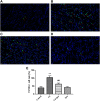
Effects of Quercetin (Que) on the rate of TUNEL-positive apoptosis in Cd-induced kidney injury rats. (A) TUNEL-positive cells in the control group 400×. (B) TUNEL-positive cells in the Cd-treated group 400×. (C) TUNEL-positive cells in the Cd + Que-treated group 400×. (D) TUNEL-positive cells in the Que-treated group 400×. The green particles are apoptotic cells, and the blue particles are normal or proliferating cells. (E) The percentage of TUNEL-positive cells in control and Cd groups within or without Que using TUNEL staining. Data were expressed as mean ± SEM. n = 3, **p < 0.01 indicates extremely significant difference compared to the control group; ## p < 0.01 indicates extremely significant difference compared to the Cd group.
FIGURE 6
FIGURE 6
Effects of Quercetin (Que) on mRNA expression of apoptosis-related genes in Cd-induced kidney injury rats. The mRNA expression of Caspase-9, Caspase-3 and Cyt-c in kidney tissue was reduced by Que. Data were expressed as mean ± SEM. n = 3, *p < 0.05, **p < 0.01 indicates a significant or extremely significant difference compared to the control group; # p < 0.05, ## p < 0.01 indicates a significant or extremely significant difference compared to the Cd group.
FIGURE 7
FIGURE 7
Effects of Quercetin (Que) on the levels of apoptosis-related proteins in Cd-induced kidney injury rats. (A) Western blotting was used to determine the amounts of Caspase-9, Caspase-3, and Cyt-c proteins, with β-Actin as a control. Quantitative analysis for Caspase-9 (B), Caspase-3 (C) and Cyt-c (D). The results reflect the mean ± SEM of three separate experiments. **p < 0.01 indicates extremely significant difference compared to the control group; # p < 0.05, ## p < 0.01 indicates a significant or extremely significant difference compared to the Cd group.

Similar articles

Cited by

References

    1. Bauer T. M., Murphy E. (2020). Role of mitochondrial calcium and the permeability transition pore in regulating cell death. Circ. Res. 126 (2), 280–293. 10.1161/CIRCRESAHA.119.316306 - DOI - PMC - PubMed
    1. Bhattacharya S., Haldar P. K. (2012). Trichosanthes dioica fruit ameliorates experimentally induced arsenic toxicity in male albino rats through the alleviation of oxidative stress. Biol. Trace Elem. Res. 148 (2), 232–241. 10.1007/s12011-012-9363-3 - DOI - PubMed
    1. Carrasco-Torres G., Monroy-Ramírez H. C., Martínez-Guerra A. A., Baltiérrez-Hoyos R., Romero-Tlalolini M., Villa-Treviño S., et al. (2017). Quercetin reverses rat liver preneoplastic lesions induced by chemical carcinogenesis. Oxid. Med. Cell. Longev. 2017, 4674918. 10.1155/2017/4674918 - DOI - PMC - PubMed
    1. Chen H. H., Chen S. P., Zheng Q. L., Nie S. P., Li W. J., Hu X. J., et al. (2018). Genistein promotes proliferation of human cervical cancer cells through estrogen receptor-mediated PI3K/Akt-NF-κB pathway. J. Cancer 9 (2), 288–295. 10.7150/jca.20499 - DOI - PMC - PubMed
    1. Choe J. Y., Park K. Y., Kim S. K. (2015). Oxidative stress by monosodium urate crystals promotes renal cell apoptosis through mitochondrial caspase-dependent pathway in human embryonic kidney 293 cells: Mechanism for urate-induced nephropathy. Apoptosis 20 (1), 38–49. 10.1007/s10495-014-1057-1 - DOI - PubMed