

Changes in vastus lateralis fibre cross-sectional area, pennation angle and fascicle length do not predict changes in muscle cross-sectional area
- PMID: 36053170
- PMCID: PMC9633374
- DOI: 10.1113/EP090666
Changes in vastus lateralis fibre cross-sectional area, pennation angle and fascicle length do not predict changes in muscle cross-sectional area
Abstract
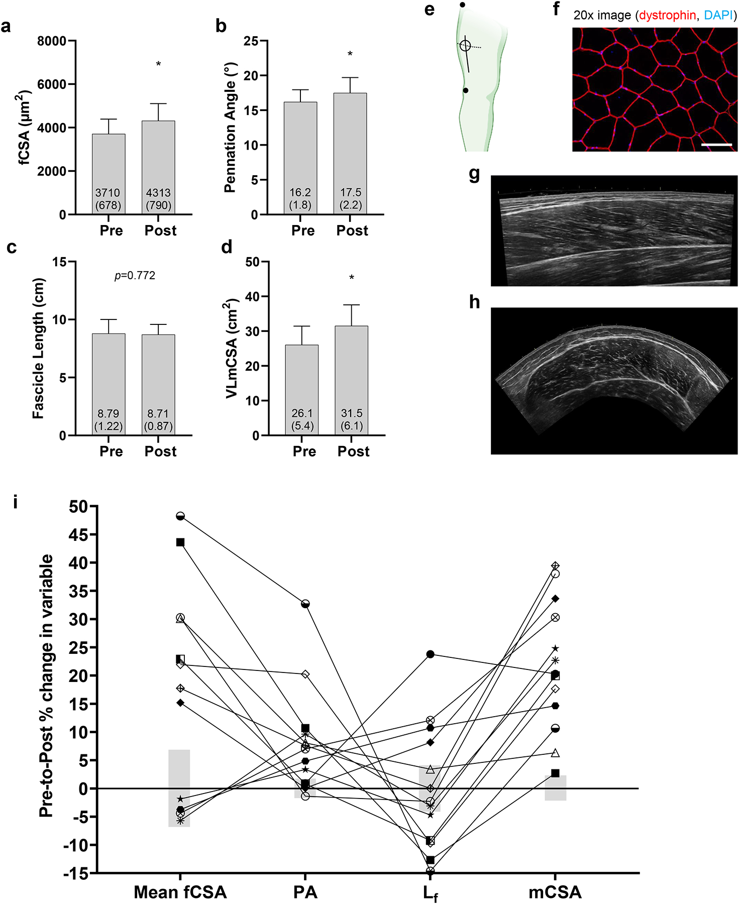
New findings: What is the central question of this study? Do changes in myofibre cross-sectional area, pennation angle and fascicle length predict vastus lateralis whole-muscle cross-sectional area changes following resistance training? What is the main finding and its importance? Changes in vastus lateralis mean myofibre cross-sectional area, fascicle length and pennation angle following a period of resistance training did not collectively predict changes in whole-muscle cross-sectional area. Despite the limited sample size in this study, these data reiterate that it remains difficult to generalize the morphological adaptations that predominantly drive tissue-level vastus lateralis muscle hypertrophy.
Abstract: Myofibre hypertrophy during resistance training (RT) poorly associates with tissue-level surrogates of hypertrophy. However, it is underappreciated that, in pennate muscle, changes in myofibre cross-sectional area (fCSA), fascicle length (Lf ) and pennation angle (PA) likely coordinate changes in whole-muscle cross-sectional area (mCSA). Therefore, we determined if changes in fCSA, PA and Lf predicted vastus lateralis (VL) mCSA changes following RT. Thirteen untrained college-aged males (23 ± 4 years old, 25.4 ± 5.2 kg/m2 ) completed 7 weeks of full-body RT (twice weekly). Right leg VL ultrasound images and biopsies were obtained prior to (PRE) and 72 h following (POST) the last training bout. Regression was used to assess if training-induced changes in mean fCSA, PA and Lf predicted VL mCSA changes. Correlations were also performed between PRE-to-POST changes in obtained variables. Mean fCSA (+18%), PA (+8%) and mCSA (+22%) increased following RT (P < 0.05), but not Lf (0.1%, P = 0.772). Changes in fCSA, Lf and PA did not collectively predict changes in mCSA (R2 = 0.282, adjusted R2 = 0.013, F3,8 = 1.050, P = 0.422). Moderate negative correlations existed for percentage changes in PA and Lf (r = -0.548, P = 0.052) and changes in fCSA and Lf (r = -0.649, P = 0.022), and all other associations were weak (|r| < 0.500). Although increases in mean fCSA, PA and VL mCSA were observed, inter-individual responses for each variable and limitations for each technique make it difficult to generalize the morphological adaptations that predominantly drive tissue-level VL muscle hypertrophy. However, the small subject pool is a significant limitation, and more research in this area is needed.
Keywords: histology; muscle; resistance training; ultrasound.
© 2022 The Authors. Experimental Physiology © 2022 The Physiological Society.
Conflict of interest statement
Competing interests
None of the authors have financial or other conflicts of interest to report regarding these data.
Figures

References
-
- Ahtiainen JP, Hoffren M, Hulmi JJ, Pietikainen M, Mero AA, Avela J & Hakkinen K (2010). Panoramic ultrasonography is a valid method to measure changes in skeletal muscle cross-sectional area. Eur J Appl Physiol 108, 273–279. - PubMed
-
- American College of Sports M, Sawka MN, Burke LM, Eichner ER, Maughan RJ, Montain SJ & Stachenfeld NS (2007). American College of Sports Medicine position stand. Exercise and fluid replacement. Med Sci Sports Exerc 39, 377–390. - PubMed
-
- Angleri V, Damas F, Phillips SM, Selistre-de-Araujo HS, Cornachione AS, Stotzer US, Santanielo N, Soligon SD, Costa LAR, Lixandrao ME, Conceicao MS, Vechin FC, Ugrinowitsch C & Libardi CA (2022). Resistance training variable manipulations are less relevant than intrinsic biology in affecting muscle fiber hypertrophy. Scand J Med Sci Sports 32, 821–832. - PubMed
-
- Baar K, Nader G & Bodine S (2006). Resistance exercise, muscle loading/unloading and the control of muscle mass. Essays Biochem 42, 61–74. - PubMed
Publication types
MeSH terms
Grants and funding
LinkOut - more resources
Full Text Sources
Miscellaneous