. 2022 Sep 20;55(18):2728-2739.
doi: 10.1021/acs.accounts.2c00458. Epub 2022 Sep 2.

The Dual Use of the Pyranine (HPTS) Fluorescent Probe: A Ground-State pH Indicator and an Excited-State Proton Transfer Probe

Affiliations

The Dual Use of the Pyranine (HPTS) Fluorescent Probe: A Ground-State pH Indicator and an Excited-State Proton Transfer Probe

Ramesh Nandi et al. Acc Chem Res. .

Abstract

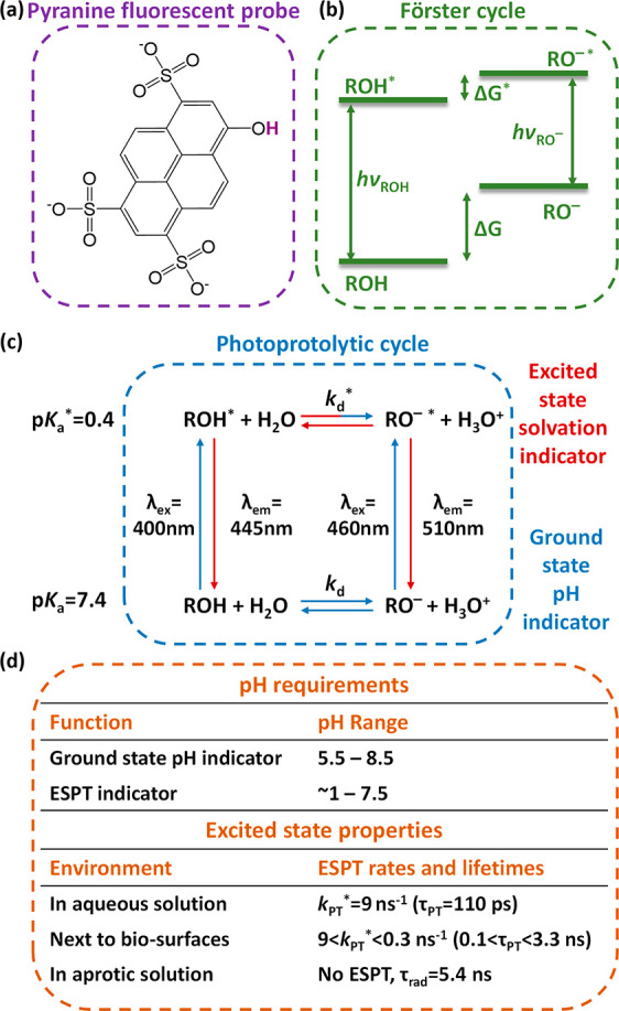
Molecular fluorescent probes are an essential experimental tool in many fields, ranging from biology to chemistry and materials science, to study the localization and other environmental properties surrounding the fluorescent probe. Thousands of different molecular fluorescent probes can be grouped into different families according to their photophysical properties. This Account focuses on a unique class of fluorescent probes that distinguishes itself from all other probes. This class is termed photoacids, which are molecules exhibiting a change in their acid-base transition between the ground and excited states, resulting in a large change in their pKa values between these two states, which is thermodynamically described using the Förster cycle. While there are many different photoacids, we focus only on pyranine, which is the most used photoacid, with pKa values of ∼7.4 and ∼0.4 for its ground and excited states, respectively. Such a difference between the pKa values is the basis for the dual use of the pyranine fluorescent probe. Furthermore, the protonated and deprotonated states of pyranine absorb and emit at different wavelengths, making it easy to focus on a specific state. Pyranine has been used for decades as a fluorescent pH indicator for physiological pH values, which is based on its acid-base equilibrium in the ground state. While the unique excited-state proton transfer (ESPT) properties of photoacids have been explored for more than a half-century, it is only recently that photoacids and especially pyranine have been used as fluorescent probes for the local environment of the probe, especially the hydration layer surrounding it and related proton diffusion properties. Such use of photoacids is based on their capability for ESPT from the photoacid to a nearby proton acceptor, which is usually, but not necessarily, water. In this Account, we detail the photophysical properties of pyranine, distinguishing between the processes in the ground state and the ones in the excited state. We further review the different utilization of pyranine for probing different properties of the environment. Our main perspective is on the emerging use of the ESPT process for deciphering the hydration layer around the probe and other parameters related to proton diffusion taking place while the molecule is in the excited state, focusing primarily on bio-related materials. Special attention is given to how to perform the experiments and, most importantly, how to interpret their results. We also briefly discuss the breadth of possibilities in making pyranine derivatives and the use of pyranine for controlling dynamic reactions.


Conflict of interest statement

The authors declare no competing financial interest.

Figures

Figure 1
Figure 1
(a) Chemical structure, (b) Förster cycle, and (c) photoprotolytic cycle of pyranine. (d) Summary of the important requirements and excited-state properties for the use of pyranine as a probe (including the ESPT rate (kPT*) and lifetime (τPT) and the pure radiative lifetime (τrad).
Figure 2
Figure 2
(a) Excitation (λem = 510 nm) and emission (λex = 400 nm) spectra of pyranine in aqueous solutions at pH 4 (solid), pH 7 (dashed), and pH 10 (dot-dashed). (b) Ratio of relative intensities (I450nm/I400nm) in the excitation spectra (λem = 510 nm) of pyranine as a function of pH. The inset shows the titration curves of pyranine in the same solutions. (c) Schematic of the use of pyranine (cyan spheres) within liposomes. Reproduced with permission from ref (14). Copyright 1978 Elsevier.
Figure 3
Figure 3
Time-resolved fluorescence decay of (a) ROH and (b) RO (the insets show the first nanosecond of the decay) and (c) steady-state fluorescence spectra of pyranine at different pH values and in methanol.
Figure 4
Figure 4
Constructed time-resolved transients of IfROH* using the SSDP software (a) at different kPT* values (ka* = 9 ns–1) and (b) at different ka* values (kPT* = 9 ns–1). The right panels are zoomed-in views of the first 150 ps.
Figure 5
Figure 5
(a) Schematic of the use of pyranine (cyan spheres) within reverse micelles, (b) fluorescence decay (on a log–log plot) of pyranine within them (where w0 is the mole ratio of water to surfactant molecules), and (c) the kPT* and ka* values extracted using an exponential fit of the decay. Reproduced from ref (45). Copyright 2007 American Chemical Society.
Figure 6
Figure 6
Time-resolved emission of pyranine in BSA fractions of (a) ≤ 0.75% and (b) ≥ 0.75%. The insets show magnifications of the first nanoseconds. (c) Schematic of the system. Reproduced with permission from ref (1). Copyright 2015 The PCCP Owner Societies.
Figure 7
Figure 7
(a) Schematic of pyranine on the surface of fibrils. (b–d) Time-resolved emission of pyranine (b) on the surface of BSA before and after thermal treatment for the 0.5% and 5% BSA samples, where only the 5% samples undergo gelation; (c) on the surface of protein mats at different wt % of water compared to pyranine in bulk water; and (d) on the surface of amyloid fibrils at different pH values. Panel (b) is reproduced with permission from ref (3). Copyright 2020 Royal Society of Chemistry. Panel (c) is from ref (59). CC BY 4.0. Panel (d) is reproduced from ref (61). Copyright 2014 American Chemical Society.
Figure 8
Figure 8
(a, b) Time-resolved emission of pyranine (a) adsorbed on cellulose in the dry state for several weight percentages of H2O added (relative to cellulose), compared to pyranine in bulk water together with the extracted parameters according to eq 1, and (b) adsorbed on cellulose and chitin in a methanolic solution, compared to pyranine in bulk methanol. (c) Schematic of the experimental systems showing the two possibilities for ESPT. Reproduced from refs (61) and (63). Copyright 2014 and 2015, respectively, American Chemical Society.
Figure 9
Figure 9
(a) Molecular structure of C12-HPTS. (b) Schematic of the ESPT processes involving the surface of the membrane. (c) Time-resolved emission of pyranine in several lipid vesicles at pH 6.5. (d) Extracted parameters according to eq 1. Reproduced from ref (2).
Figure 10
Figure 10
Various reported derivatives of pyranine photoacids and their unique features.

Similar articles

Cited by

References

    1. Amdursky N. Photoacids as a new fluorescence tool for tracking structural transitions of proteins: following the concentration-induced transition of bovine serum albumin. Phys. Chem. Chem. Phys. 2015, 17, 32023–32032. 10.1039/C5CP05548B. - DOI - PubMed
    1. Amdursky N.; Lin Y.; Aho N.; Groenhof G. Exploring fast proton transfer events associated with lateral proton diffusion on the surface of membranes. Proc. Natl. Acad. Sci. U. S. A. 2019, 116, 2443–2451. 10.1073/pnas.1812351116. - DOI - PMC - PubMed
    1. Nandi R.; Yucknovsky A.; Mazo M. M.; Amdursky N. Exploring the inner environment of protein hydrogels with fluorescence spectroscopy towards understanding their drug delivery capabilities. J. Mater. Chem. B 2020, 8, 6964–6974. 10.1039/D0TB00818D. - DOI - PubMed
    1. Burnstine-Townley A.; Mondal S.; Agam Y.; Nandi R.; Amdursky N. Light-modulated cationic and anionic transport across protein biopolymers. Angew. Chem., Int. Ed. 2021, 60, 24676–24685. 10.1002/anie.202111024. - DOI - PubMed
    1. Amdursky N.; Erez Y.; Huppert D. Molecular Rotors: What Lies Behind the High Sensitivity of the Thioflavin-T Fluorescent Marker. Acc. Chem. Res. 2012, 45, 1548–1557. 10.1021/ar300053p. - DOI - PubMed

Publication types