. 2022 Oct;33(19-20):1091-1100.
doi: 10.1089/hum.2022.132.

Urocortin 2 Gene Transfer for Systolic and Diastolic Dysfunction Due to Chronically Increased Left Ventricular Pressure

Affiliations

Urocortin 2 Gene Transfer for Systolic and Diastolic Dysfunction Due to Chronically Increased Left Ventricular Pressure

N Chin Lai et al. Hum Gene Ther. 2022 Oct.

Abstract

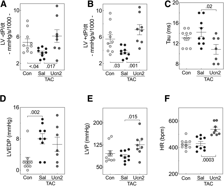
We used transverse aortic constriction (TAC) in mice to test the hypothesis that urocortin 2 (Ucn2) gene transfer would increase left ventricular (LV) systolic and diastolic function in the pressure-stressed LV. Three groups were studied: (1) control mice (no TAC); (2) mice that received saline 6 weeks after TAC; and (3) mice that received Ucn2 gene transfer 6 weeks after TAC, using adeno-associated virus 8 encoding murine Ucn2 (AAV8.mUcn2; 2 × 10¹³ genome copies (gc)/kg, i.v. per mouse). Echocardiography was performed 6 and 12 weeks after TAC. In terminal studies 12 weeks after TAC, rates of LV pressure development and decay and Tau were measured, and LV cardiac myocytes (CMs) were isolated and cytosolic Ca2+ transients and sarcomere shortening rates recorded. Reverse transcription polymerase chain reaction and immunoblotting were used to measure key proteins in LV samples. A CM cell line (HL-1) was used to explore mechanisms. Concentric LV hypertrophy was evident on echocardiography 6 weeks after TAC. Twelve weeks after TAC, LV ejection fraction (EF) was higher in mice that received Ucn2 gene transfer (TAC-saline: 65% ± 3%; TAC-Ucn2: 75% ± 2%; p = 0.01), as was LV peak +dP/dt (1.9-fold increase; p = 0.001) and LV peak -dP/dt (1.7-fold increase; p = 0.017). Tau was more rapid (23% reduction, p = 0.02), indicating improved diastolic function. The peak rates of sarcomere shortening (p = 0.002) and lengthening (p = 0.002) were higher in CMs from TAC-Ucn2 mice, and Tau was reduced (p = 0.001). LV (Ser-16) phosphorylation of phospholamban (PLB) was increased in TAC-Ucn2 mice (p = 0.025), and also was increased in HL-1 cells treated with angiotensin II to induce hypertrophy and incubated with Ucn2 peptide (p = 0.001). Ucn2 gene transfer in TAC-induced heart failure with preserved ejection fraction increased cardiac function in the intact LV and provided corresponding benefits in CMs isolated from study animals, including increased myofilament Ca2+ sensitivity during contraction. The mechanism includes enhanced CM Ca2+ handling associated with increased (Ser-16)-PLB.

Keywords: HL-1 cell line; TAC; Tau; gene transfer.


Conflict of interest statement

Dr. Hammond is a founder, board member, and unpaid consultant for Renova Therapeutics. Renova played no financial, intellectual, or editorial role in the studies. None of the other authors has disclosures.

Figures

Figure 1.
Figure 1.
Study design and Ucn2 expression. (A) Mice (n = 54) underwent TAC; a control group (n = 18) did not receive TAC. Six weeks later, 21 mice received Ucn2 gene transfer (AAV8.mUcn2, 2 × 1013 gc/kg, i.v.) and 29 mice received saline, i.v. The control group received no intervention. Six weeks later, mice underwent the final studies depicted. (B) Mice that received Ucn2 gene transfer (TAC-Ucn2) showed a mean 20-fold increase in plasma Ucn2 versus TAC-saline animals (p < 0.003). (C, D) Ucn2 mRNA expression was increased in the liver (>85,000-fold control; p < 0.004) and LV (>900-fold control, p < 0.04). p Values from Student's t-test (unpaired and two tailed). Data denote mean value ± SE. IB, immunoblotting; LV, left ventricular; SE, standard error; TAC, transverse aortic constriction; Ucn2 GT, urocortin 2 gene transfer.
Figure 2.
Figure 2.
Effects of Ucn2 gene transfer on LV function. Mice underwent TAC, were randomized 6 weeks later to receive i.v. saline or Ucn2 gene transfer (AAV8.mUcn2, 2 × 1013 gc/kg, i.v.), and were studied 6 weeks after randomization. A control group (age- and strain-matched) did not undergo TAC (see Fig. 1A). Data were obtained from anesthetized mice using Millar pressure catheters in terminal studies. (A) TAC was associated with reduced LV peak −dP/dt (p < 0.04 vs. control). Mice that received Ucn2 gene transfer showed a higher peak rate of LV pressure decline (−dP/dt; p = 0.017), which was similar to normal mice. (B) TAC was associated with reduced LV peak +dP/dt (p = 0.03 vs. control). Mice that received Ucn2 gene transfer showed a higher rate of LV pressure rise, which was similar to normal mice (+dP/dt; p = 0.001). (C) TAC-Ucn2 mice showed more rapid Tau (p = 0.02; time constant of LV pressure decay), indicating increased LV diastolic function. (D) TAC was associated with increased LVEDP (p = 0.002). (E) TAC-Ucn2 mice showed increased LVP (p = 0.015). (F) TAC-Ucn2 mice showed a higher HR during anesthesia (p = 0.0003). p Values from Student's t-test (unpaired and two tailed). Data denote mean values ± SE. HR, heart rate; LVEDP, LV end-diastolic pressure; LVP, LV developed pressure.
Figure 3.
Figure 3.
Ca2+ transients and sarcomere shortening. (A) Representative Ca2+ transients from each group. (B) Peak rates of Ca2+ transients were lower in CMs from TAC-saline mice versus control (B, p = 0.03) and tended to be higher in CMs from TAC-Ucn2 versus TAC-saline mice (B, p = 0.06). (C–G) Sarcomere shortening. (C) Representative sarcomere shortening from each group. (D) Sarcomere fractional shortening was higher in CMs from TAC-Ucn2 versus TAC-saline mice (p < 0.02). (E) TAC was associated with prolonged Tau, the time constant of sarcomere relaxation (p = 0.04 vs. control), indicating impaired relaxation. In contrast, Tau was more rapid in CM from TAC-Ucn2 versus TAC-saline mice (p = 0.001), indicating enhanced diastolic function. (F) The peak rate of sarcomere shortening (+dL/dt) was more rapid in CM from TAC-Ucn2 versus TAC-saline mice (F, p = 0.002). (G) Likewise, the peak rate of sarcomere lengthening (−dL/dt) was more rapid in CM from TAC-Ucn2 versus TAC-saline mice (G, p = 0.001). (H) Myofilament sensitivity. Myofilament sensitivity to Ca2+ was analyzed from Ca2+ transients and the coinciding sarcomere shortening and displayed as mean phase loops from CMs from each group. Arrows indicate loop direction. The loop from CMs from TAC-Ucn2 12 weeks after TAC shows a leftward shift compared with the TAC-saline group, indicating increased myofilament sensitivity to cytosolic Ca2+. (I) During contraction, EC50 for cytosolic Ca2+ (Indo-1 ratio) was reduced in CM from TAC-Ucn2 mice during the contraction phase (p = 0.012), indicating increased myofilament Ca2+ sensitivity. (J) There were no group differences during the relaxation phase. p Values from Student's t-test (unpaired and two tailed). Data denote mean values ± SE. CM, cardiac myocyte.
Figure 4.
Figure 4.
LV immunoblotting. (A) Neither TAC nor Ucn2 gene transfer altered LV SERCA2a content. (B) LV PLB phosphorylation at Ser16 was restored to normal levels by Ucn2 gene transfer (p = 0.025). (C) LV total PLB was higher after TAC (p = 0.001), but there was no difference between TAC-saline versus TAC-Ucn2. (D) Cultured HL-1 cells were exposed to AngII (100 nM, 24 h) to induce hypertrophy, documented by reduced phosphorylation at Thr56 of eEF2. The addition of Ucn2 peptide (100 nM, 15 min) increased PLB phosphorylation at Ser16, both in the absence (p = 0.0016) and presence of AngII (p = 0.001). Data displayed are normalized to vinculin (A) or GAPDH (B–D). p Values from Student's t-test. Data denote mean value ± SE. AngII, angiotensin II; Con, control; eEF2, eukaryotic elongation factor; GAPDH, glyceraldehyde-3-phosphate dehydrogenase; PLB, phospholamban; Sal, saline; Uc, Ucn2.

Similar articles

References

    1. Lai NC, Gao MH, Giamouridis D, et al. . Intravenous AAV8 encoding urocortin-2 increases function of the failing heart in mice. Hum Gene Ther 2015;26(6):347–356; doi: 10.1089/hum.2014.157 - DOI - PMC - PubMed
    1. Giamouridis D, Gao MH, Lai NC, et al. . Urocortin 2 gene transfer improves heart function in aged mice. Mol Ther 2020;28(1):180–188; doi: 10.1016/j.ymthe.2019.10.003 - DOI - PMC - PubMed
    1. Miura T, Bhargava V, Guth BD, et al. . Increased afterload intensifies asynchronous wall motion and impairs ventricular relaxation. J Appl Physiol 1993;75(1):389–396; doi: 10.1152/jappl.1993.75.1.389 - DOI - PubMed
    1. Barrick CJ, Rojas M, Schoonhoven R, et al. . Cardiac response to pressure overload in 129S1/SvImJ and C57BL/6J mice: Temporal- and background-dependent development of concentric left ventricular hypertrophy. Am J Physiol Heart Circ Physiol 2007;292(5):H2119–H2130; doi: 10.1152/ajpheart.00816.2006 - DOI - PubMed
    1. Valero-Munoz M, Backman W, Sam F. Murine models of heart failure with preserved ejection fraction: A “fishing expedition.” JACC Basic Transl Sci 2017;2(6):770–789; doi: 10.1016/j.jacbts.2017.07.013 - DOI - PMC - PubMed

Publication types