Review
. 2023 Mar;38(7-9):550-580.
doi: 10.1089/ars.2022.0110. Epub 2022 Oct 12.

The Interplay Between Autophagy and Regulated Necrosis

Affiliations
Review

The Interplay Between Autophagy and Regulated Necrosis

Lei Zhang et al. Antioxid Redox Signal. 2023 Mar.

Abstract

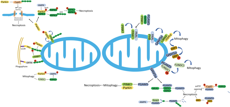
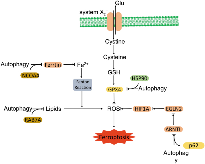
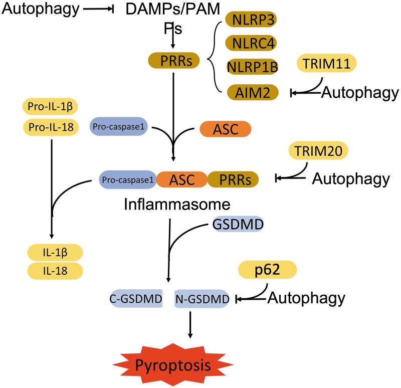
Significance: Autophagy is critical to cellular homeostasis. Emergence of the concept of regulated necrosis, such as necroptosis, ferroptosis, pyroptosis, and mitochondrial membrane-permeability transition (MPT)-derived necrosis, has revolutionized the research into necrosis. Both altered autophagy and regulated necrosis contribute to major human diseases. Recent studies reveal an intricate interplay between autophagy and regulated necrosis. Understanding the interplay at the molecular level will provide new insights into the pathophysiology of related diseases. Recent Advances: Among the three forms of autophagy, macroautophagy is better studied for its crosstalk with regulated necrosis. Macroautophagy seemingly can either antagonize or promote regulated necrosis, depending upon the form of regulated necrosis, the type of cells or stimuli, and other cellular contexts. This review will critically analyze recent advances in the molecular mechanisms governing the intricate dialogues between macroautophagy and main forms of regulated necrosis. Critical Issues: The dual roles of autophagy, either pro-survival or pro-death characteristics, intricate the mechanistic relationship between autophagy and regulated necrosis at molecular level in various pathological conditions. Meanwhile, key components of regulated necrosis are also involved in the regulation of autophagy, which further complicates the interrelationship. Future Directions: Resolving the controversies over causation between altered autophagy and a specific form of regulated necrosis requires approaches that are more definitive, where rigorous evaluation of autophagic flux and the development of more reliable and specific methods to quantify each form of necrosis will be essential. The relationship between chaperone-mediated autophagy or microautophagy and regulated necrosis remains largely unstudied. Antioxid. Redox Signal. 38, 550-580.

Keywords: autophagy; ferroptosis; mitochondrial membrane permeability transition; mitophagy; necroptosis; pyroptosis.


Conflict of interest statement

The authors declare that there is no conflict of interest.

Figures

FIG. 1.
FIG. 1.
Overview of molecular mechanism of autophagy. AMPK-TSC1/2 signaling positively or PI3K-AKT signaling negatively regulates the activation of autophagy through inhibiting or activating mTORC1, respectively. Activation of the ULK1 complex and the PtdIns3K complex induces the nucleation of phagophore. The following elongation of phagophore involves the ATG12–ATG5-ATG16 complex and LC3-II that is generated from LC3-I through lipidation by the cooperation of ATG4 and ATG7. Eventually, the expanding membrane closes to engulf the cargoes, forming the autophagosome. Then, the autophagosome fuses with lysosomes and thereby forms autolysosome. The cargo and the inner membrane of the autophagosome are degraded and recycled by the lysosomal enzymes. AKT, kinase AKT, also known as protein kinase B; AMPK, 5′ AMP-activated protein kinase; ATG, autophagy related gene; LC3, microtubule-associated protein 1 light chain 3; mTORC1, mechanistic target of rapamycin complex 1; PI3K, phosphatidylinositol 3 kinase; TSC, tuberous sclerosis; ULK, Unc-51 like autophagy activating kinase.
FIG. 2.
FIG. 2.
Overview of the canonical pathway to necroptosis. The binding of TNFα to TNFR1 recruits complex I (RIPK1, TRADD, cIAP1, TRAF2/5). In complex I, RIPK1 is rapidly polyubiquitinated by cIAP1/2 mediated K63-linked ubiquitin chains and LUBAC mediated M1-linked ubiquitin chains. K63 ubiquitin chains recruit TAK1, leading to phosphorylation of RIPK1 at Ser321. The phosphorylation may determine the cell fate through (1) activating IKK to facilitate NF-κB dependent cell survival or (2) promoting the necroptosis. CYLD deubiquitinates M1-ubiquitin chains of RIPK1, which promotes the formation of complex IIb/necrosome. The complex IIb subsequently phosphorylates MLKL and promotes the oligomerization of MLKL at the plasma membrane to induce necroptotic cell death. A20 promotes the deubiquitination of K63-ubiquitin chain of RIPK1, and the deubiquitinated RIPK1 forms the complex IIa with FADD, TRADD, and Caspase 8. The complex IIa initiates apoptosis. Inhibition of Caspase 8 switches the apoptosis to RIPK1 dependent necroptosis. cIAP, cellular inhibitor of apoptosis protein; CYLD, cylindromatosis; FADD, FAS-associated death domain protein; IKK, IκB kinase; LUBAC, linear ubiquitination assembly complex; M1, Met1; MLKL, mixed lineage kinase domain like psudokinase; NF-κB, nuclear factor kappa light chain enhancer of activated B cell; RIPK, receptor interacting protein kinase; TAK1, TGFβ activating kinase 1; TNFα, tumor necrosis factor alpha; TNFR, TNF receptor; TRADD, TNFR-associated death domain; TRAF, TNF receptor-associated factor.
FIG. 3.
FIG. 3.
Autophagy regulates necroptosis. Autophagy either inhibits (illustrated by red squares) or promotes (illustrated by blue squares) necroptosis. (a) ULK1 phosphorylates RIPK1 at Ser357, which inhibits the formation of necrosome. (b) Beclin1 disrupts the formation of MLKL oligomerization. (c) Autophagy promotes the degradation of key components of the necroptotic pathway. (d) Autophagy facilitates the degradation of autophagic receptor, p62, the molecule that promotes the formation of necrosome. (e) ATG5 promotes the formation of necrosome directly or indirectly by enhancing the interaction of RIPKs with p62. (f) Autophagy promotes necroptosis by degradation of inhibitory molecules of necroptosis, cIAP1/2. (g) Autophagy promotes necroptosis by degradation of caspase 8, which blocks the cleavage of RIPK1.
FIG. 4.
FIG. 4.
Necroptosis regulates autophagy. Key components of necroptosis either promote or inhibit autophagy. RIPK1 and RIPK3 promote the activation of AMPK, which promotes the activation of autophagy through the AMPK/TSC/mTORC1 signaling axis. RIPK3 phosphorylates ULK1 to facilitate alternative autophagy. On the other hand, RIPK1 inhibits autophagy through activation of NF-κB. RIPK1 and RIPK3 inhibit the fusion of autophagosomes with lysosomes by disrupting the interactions of SNARE complexes. MLKL activates the PI3K/AKT/mTOR signaling pathway to inhibit autophagy. MLKL also damages the membrane integrity of autophagosomes and disrupts the autophagic flux. mTOR, mechanistic target of rapamycin; SNARE, soluble N-ethylmaleimide-sensitive factor attachment protein receptor.
FIG. 5.
FIG. 5.
Crosstalk between mitophagy and necroptosis. Components of mitophagy and necroptosis intertwine in the crosstalk between mitophagy and necroptosis. PINK1 and Parkin coordinate to initiate mitophagy through forming phosphorylated ubiquitin chains in response to mytophagy stimuli. Parkin inhibits necroptosis through disruption of necrosome formation and CYPD-dependent MPT pore opening. PGAM5, as another key component, promotes mitophagy through stabilizing PINK1 and dephosphorylating DRP1 and FUNDC1. As a substrate of RIPK1 and RIPK3, PGAM5's phosphorylation increases its phosphatase activity, which promotes necroptosis through dephosphorylation of DRP1 and the enhanced activation of PGAM5-CYPD signaling. Necroptotic factor RIPK1 promotes mitophagy through stabilizing PINK1. RIPK3 negatively regulates mitophagy through inactivation of FUNDC1 and blockade of the AMPK-dependent activation of Parkin. CYPD, Cyclophilin D; DRP1, dynamin related protein 1; FUNDC1, FUN14 domain-containing protein 1; MPT, mitochondrial membrane-permeability transition; PGAM, phosphoglycerate mutase; PINK1, phosphatase and tensin homolog (PTEN)-induced putative kinase 1.
FIG. 6.
FIG. 6.
Autophagy promotes ferroptosis. Autophagy facilitates degradation of ARNTL (clockophagy) and promotes ferroptosis through the EGLN2-HIF1α axis. With the assistance of NCOA4, autophagy promotes the degradation of ferritin (ferrotinophagy), which releases ferrous iron to facilitate lipid peroxidation and ferroptosis. HSP90-mediated autophagy promotes the removal of GPX4 and accumulates ROS, which facilitates the ferroptosis. Autophagy-dependent lipid droplet degradation facilitates lipid peroxidation-mediated ferroptosis. ARNTL, aryl hydrocarbon receptor nuclear translocator-like; EGLN2, egl nine homolog 2; GPX4, glutathione peroxidase 4; HIF1α, hypoxia inducible factor 1α; HSP, heat shock protein; NCOA4, nuclear receptor coactivator 4; ROS, reactive oxygen species.
FIG. 7.
FIG. 7.
Autophagy antagonizes pyroptosis. Autophagy eliminates DAMPs and PAMPs to block the formation of inflammasomes, which, in turn, inhibits the initiation of pyroptosis. In addition, autophagy promotes the degradation of key components of inflammasomes, including AIM2 (TRIM11-dependent degradation), NLRP3, NLRP1, and pro–caspase-1 (TRIM20-dependent degradation), as well as executor N-GSDMD (p62-dependent degradation). The dysfunctional inflammasome blocks the initiation of pyroptosis. AIM2, absent in melanoma 2; DAMP, danger-associated molecular pattern; N-GSDMD, N-terminus of GSDMD; NLRP3, NLR family pyrin domain containing 3; PAMP, pathogen-associated molecular pattern; TRIM11, tripartite motif 11.

Similar articles

Cited by

References

    1. Abe K, Yano T, Tanno M, et al. . mTORC1 inhibition attenuates necroptosis through RIP1 inhibition-mediated TFEB activation. Biochim Biophys Acta Mol Basis Dis 2019;1865(12):165552; doi: 10.1016/j.bbadis.2019.165552 - DOI - PubMed
    1. Ajoolabady A, Aslkhodapasandhokmabad H, Libby P, et al. . Ferritinophagy and ferroptosis in the management of metabolic diseases. Trends Endocrinol Metab 2021;32(7):444–462; doi: 10.1016/j.tem.2021.04.010 - DOI - PubMed
    1. Almanza A, Carlesso A, Chintha C, et al. . Endoplasmic reticulum stress signalling—From basic mechanisms to clinical applications. FEBS J 2019;286(2):241–278; doi: 10.1111/febs.14608 - DOI - PMC - PubMed
    1. Altman AM, Miller MJ, Mahmud J, et al. . Human cytomegalovirus-induced autophagy prevents necroptosis of infected monocytes. J Virol 2020;94(22):e01022-20; doi: 10.1128/JVI.01022-20 - DOI - PMC - PubMed
    1. Amarante-Mendes GP, Adjemian S, Branco LM, et al. . Pattern recognition receptors and the host cell death molecular machinery. Front Immunol 2018;9:2379; doi: 10.3389/fimmu.2018.02379 - DOI - PMC - PubMed

Publication types

LinkOut - more resources