

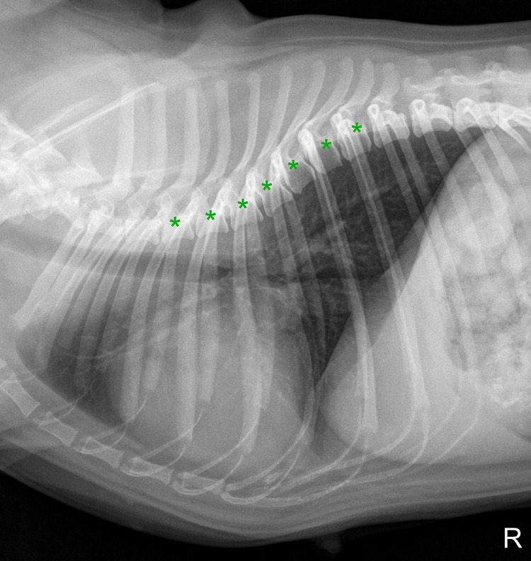
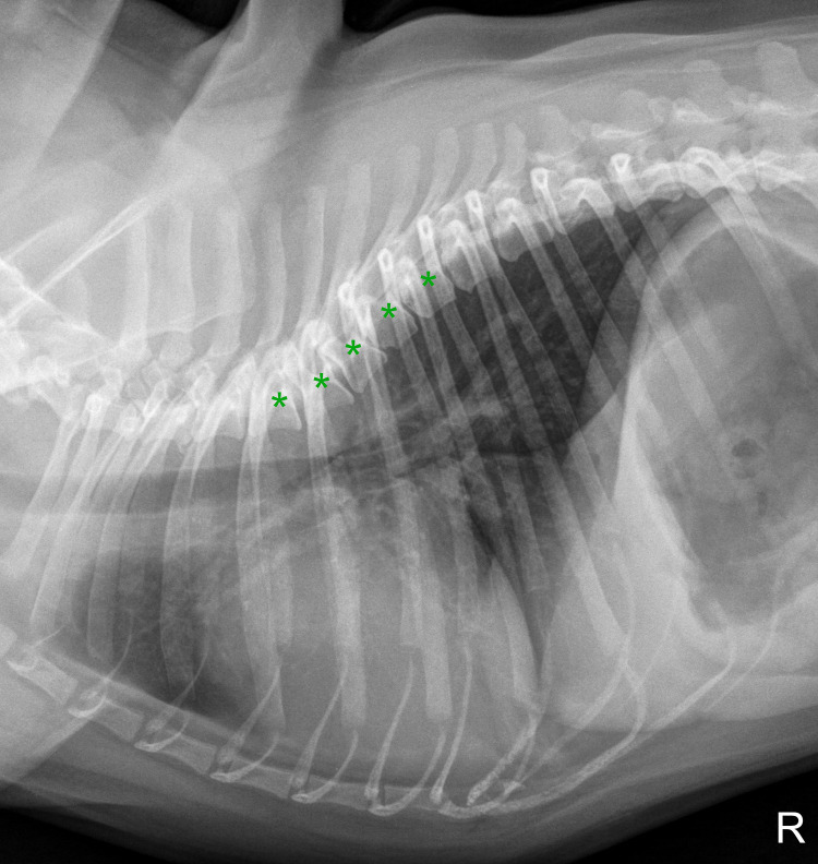
Breed-specific values for vertebral heart score (VHS), vertebral left atrial size (VLAS), and radiographic left atrial dimension (RLAD) in pugs without cardiac disease, and their relationship to Brachycephalic Obstructive Airway Syndrome (BOAS)
- PMID: 36054125
- PMCID: PMC9439199
- DOI: 10.1371/journal.pone.0274085
Breed-specific values for vertebral heart score (VHS), vertebral left atrial size (VLAS), and radiographic left atrial dimension (RLAD) in pugs without cardiac disease, and their relationship to Brachycephalic Obstructive Airway Syndrome (BOAS)
Abstract
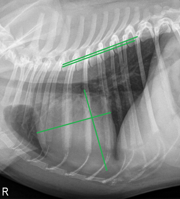
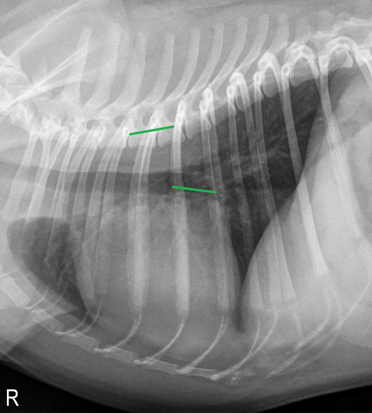
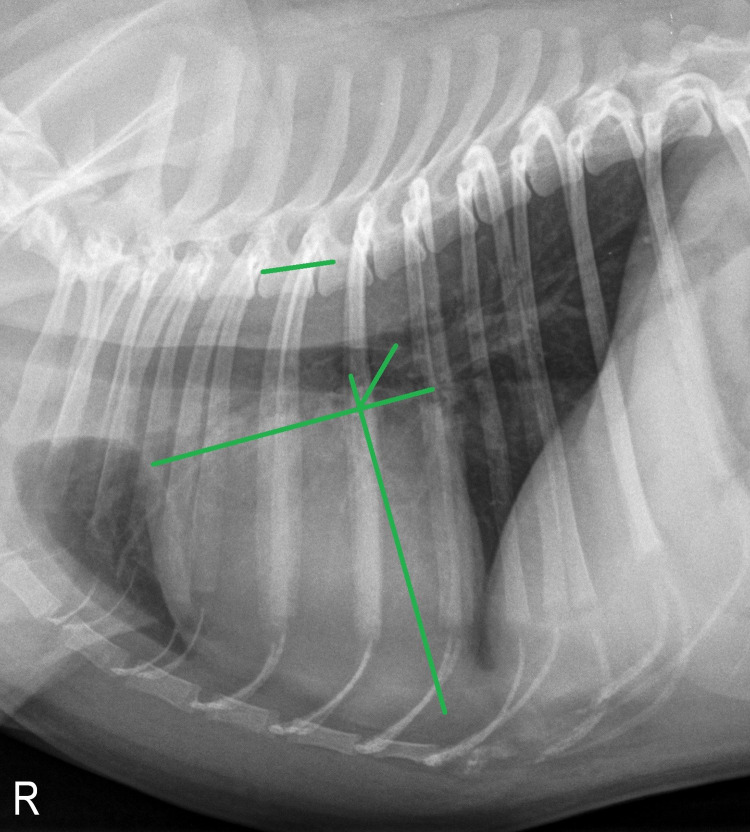
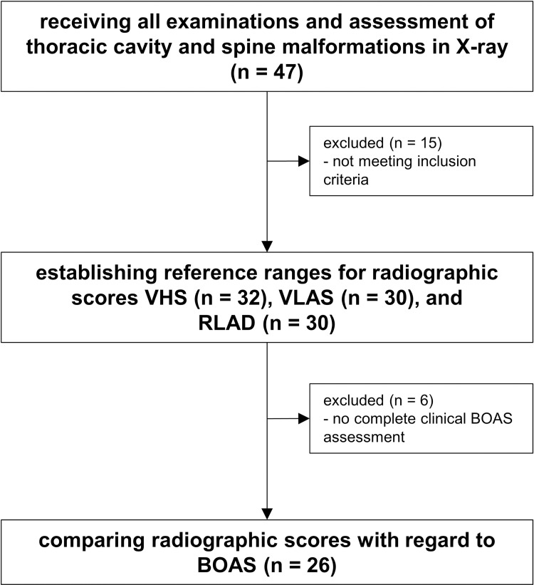
This prospective study aims to establish reference ranges for vertebral heart score (VHS), vertebral left atrial size (VLAS), and radiographic left atrial dimension (RLAD) in pugs. The impact of clinical severity of Brachycephalic Obstructive Airway Syndrome (BOAS), gender, body condition score, and body weight on VHS, VLAS, and RLAD were investigated. Intra- and interobserver correlation was determined. Correlation of radiographic scores to echocardiographic left atrial dimension was inspected. Additionally, for VLAS and RLAD, correlation to VHS was examined. Additionally, an assessment of thoracic and vertebral malformations was performed. Forty-seven privately owned pugs underwent physical examination, echocardiography, and thoracic radiography to determine cardiac health. Thirty-two pugs were eligible for establishing reference ranges for VHS in right lateral radiographs, which was 11.25 ± 0.62 (95% range, 10.1-12.8). Reference ranges for VHS in left lateral, and for VLAS and RLAD in right lateral radiograph were determined in 30 pugs. The VHS in left lateral radiograph was 11.01 ± 0.70 (95% range, 9.4-12.6), VLAS was 1.96 ± 0.38 (95% range, 1.1-2.8), and RLAD was 1.59 ± 0.34 (95% range, 0.7-2.4). Clinical severity of BOAS did not show any impact on radiographic measurements. For VLAS, a significant correlation to VHS was detected by all observers. No other variables had a consistent influence on the radiographic scores given by all observers. Interobserver agreement was almost perfect for VHS (0.89 on right lateral and 0.91 on left lateral image), moderate for VLAS (0.49), and fair for RLAD (0.22). More than one third of the entire study population (18 of 47 pugs) showed at least one thoracic cavity or spine abnormality, often leading to considerable changes in vertebral body shape and size.
Conflict of interest statement
The authors have declared that no competing interests exist.
Figures
References
-
- Malcolm EL, Visser LC, Phillips KL, Johnson LR. Diagnostic value of vertebral left atrial size as determined from thoracic radiographs for assessment of left atrial size in dogs with myxomatous mitral valve disease. J Am Vet Med Assoc. 2018;253(8):1038–45. doi: 10.2460/javma.253.8.1038 . - DOI - PubMed
Publication types
MeSH terms
LinkOut - more resources
Full Text Sources
Medical
