

Association of ictal imaging changes in status epilepticus and neurological deterioration
- PMID: 36054260
- PMCID: PMC9826342
- DOI: 10.1111/epi.17404
Association of ictal imaging changes in status epilepticus and neurological deterioration
Abstract
Objective: In patients with status epilepticus (SE), the clinical significance of ictal changes on magnetic resonance imaging (MRI) is insufficiently understood. We here studied whether the presence of ictal MRI changes was associated with neurological deterioration at discharge.
Methods: The retrospective cohort comprised all identifiable patients treated at Odense University Hospital in the period 2008-2017. All amenable MRIs were systemically screened for ictal changes. Patient demographics, electroencephalography, seizure characteristics, treatment, and SE duration were assessed. Neurological status was estimated before and after SE. The predefined endpoint was the association of neurological deterioration and ictal MRI changes.
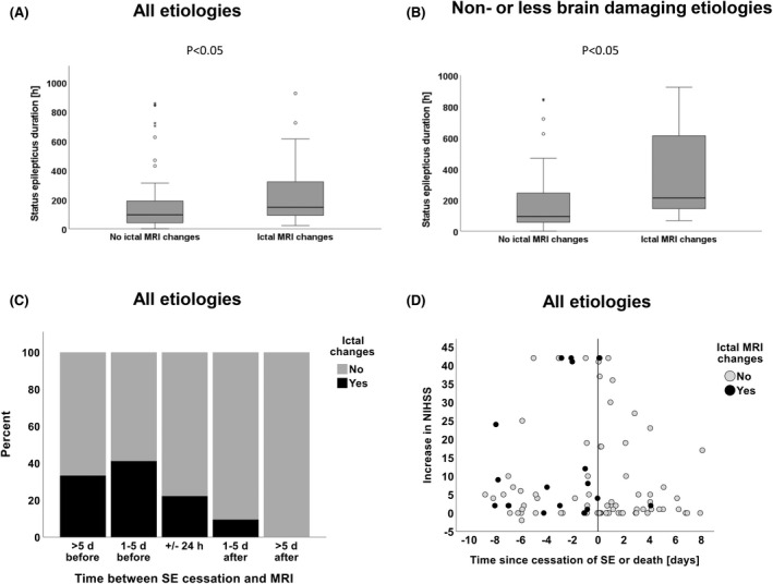
Results: Of 261 eligible patients, 101 received at least one MRI during SE or within 7 days after cessation; 43.6% (44/101) had SE due to non- or less brain-damaging etiologies. Patients who received MRI had a longer duration of SE, less frequently had a history of epilepsy, and were more likely to have SE due to unknown causes. Basic characteristics (including electroencephalographic features defined by the Salzburg criteria) did not differ between patients with (n = 20) and without (n = 81) ictal MRI changes. Timing of MRI was important; postictal changes were rare within the first 24 h and hardly seen >5 days after cessation of SE. Ictal MRI changes were associated with a higher risk of neurological deterioration at discharge irrespective of etiology. Furthermore, they were associated with a longer duration of SE and higher long-term mortality that reached statistical significance in patients with non- or less brain-damaging etiologies.
Significance: In this retrospective cohort, ictal changes on MRI were associated with a higher risk of neurological deterioration at discharge and, possibly, with a longer duration of SE and poorer survival.
Keywords: DWI; duration; ictal MRI changes; imaging; long-term outcome; status epilepticus.
© 2022 The Authors. Epilepsia published by Wiley Periodicals LLC on behalf of International League Against Epilepsy.
Conflict of interest statement
C.P.B. has received honoraria from UCB, Eisai, and Arvelle. T.K. has received honoraria from UCB. The other authors do not report possible conflicts of interest.
Figures


References
-
- Williams JA, Bede P, Doherty CP. An exploration of the spectrum of peri‐ictal MRI change; a comprehensive literature review. Seizure. 2017;50:19–32. - PubMed
-
- Kramer R, Lüders H, Lesser R, Weinstein M, Dinner D, Morris H, et al. Transient focal abnormalities of neuroimaging studies during focal status epilepticus. Epilepsia. 1987;28:528–32. - PubMed
-
- Chatzikonstantinou A, Gass A, Förster A, Hennerici MG, Szabo K. Features of acute DWI abnormalities related to status epilepticus. Epilepsy Res. 2011;97:45–51. - PubMed
-
- Raghavendra S, Ashalatha R, Krishnamoorthy T, Kesavadas C, Thomas S, Radhakrishnan K. Reversible periictal MRI abnormalities: clinical correlates and long‐term outcome in 12 patients. Epilepsy Res. 2007;73:129–36. - PubMed