Review
. 2022 Nov 30:56:279-314.
doi: 10.1146/annurev-genet-072820-034559. Epub 2022 Sep 2.

The Four Causes: The Functional Architecture of Centromeres and Kinetochores

Affiliations
Review

The Four Causes: The Functional Architecture of Centromeres and Kinetochores

Andrew D McAinsh et al. Annu Rev Genet. .

Abstract

Kinetochores are molecular machines that power chromosome segregation during the mitotic and meiotic cell divisions of all eukaryotes. Aristotle explains how we think we have knowledge of a thing only when we have grasped its cause. In our case, to gain understanding of the kinetochore, the four causes correspond to questions that we must ask: (a) What are the constituent parts, (b) how does it assemble, (c) what is the structure and arrangement, and (d) what is the function? Here we outline the current blueprint for the assembly of a kinetochore, how functions are mapped onto this architecture, and how this is shaped by the underlying pericentromeric chromatin. The view of the kinetochore that we present is possible because an almost complete parts list of the kinetochore is now available alongside recent advances using in vitro reconstitution, structural biology, and genomics. In many organisms, each kinetochore binds to multiple microtubules, and we propose a model for how this ensemble-level architecture is organized, drawing on key insights from the simple one microtubule-one kinetochore setup in budding yeast and innovations that enable meiotic chromosome segregation.

Keywords: cell division; centromere; chromatin; chromosome; kinetochore; meiosis; microtubule; mitosis; mitotic spindle.


Figures

Figure 1
Figure 1. Geometries of chromosome segregation during mitosis and meiosis.
(a) Top left: In mitosis the replicated chromosomes (sister chromatids - blue) are bioriented with sister kinetochores (red) in a back-to-back geometry and embedded in pericentromeric chromatin domain (grey). Sister chromatids are physically held together by cohesin molecules which trap the two DNA stands (black circles). The plus-end of spindle microtubules (green; either singular in budding yeast or multiple in animal cells) are embedded in the kinetochore with their minus-ends projecting towards the centrosomes (human) or spindle pole bodies (budding yeast). Pulling forces generated by kinetochore microtubule attachments pull sister chromatids apart in anaphase once cohesin is cleaved (on satisfaction of the spindle assembly checkpoint). Top Right: Mitotic spindle in a human cell (kinetochores red and microtubules (green) compared to budding yeast (kinetochores green and spindle pole bodies in red). In yeast the 32 sister kinetochores form two clusters along the spindle axis, which is ~1 μm in length. This is similar to distance between two sister kinetochores in humans. In humans the sister kinetochores are aligned along the spindle axis. (b) In meiosis I, replicated maternal and paternal (homologous) chromosomes are physically connected are a result of crossover recombination, which generates chiasmata, together with sister chromatid cohesion distal to the chiasmata. Sister kinetochores are attached to microtubules from the same pole and are said to be co-oriented. An anaphase I, cohesin is cleaved only on chromosomes arms (pericentromeric cohesin is protected from cleavage by shugoshin-PP2A; reviewed in (158)) which resolves chiasma and allows homologous chromosomes to segregate to opposite poles. In meiosis II, sister kinetochores biorient and the pericentromeric cohesin resists the pulling forces from microtubules. During anaphase II, pericentromeric cohesin is cleaved and sister kinetochores segregate to opposite poles.
Figure 2
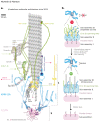
Figure 2. Molecular architecture of the kinetochore
(a) Architecture of a single microtubule-kinetochore attachment site. For clarity only one CCAN (pink) and the associated molecules is shown (see Fig. 3 for extension of models to multi-subunit kinetochores). All molecules are drawn to scale based on known structural biology, length of coiled-coil sequences or length of disordered regions. The relative position of molecules is informed by the measured Euclidian distances between the average positions of two labelled proteins in a population of kinetochores (see (213)) and/or known binding interfaces. Key features: Red circles donate known contact points between a protein and the microtubule. Flexible linkers connect CCAN to KMN (1) extended coiled-coil elements span subassembly II to sub-assembly III (CenpF, CenpE, Mad1). Detachment of microtubules triggers a switch in composition and architecture: SAC factors (yellow) including Mad1:Mad2 load on Bub1-Bub3 that are bound on the Knl1 phospho-domain (black dots) which causes rearrangement of NDC80C as they jack-knife and loose order (2). Other factors that load or leave are designated by green and red dotted arrows respectively. Not all factors are shown. Scale bar = 10 nm. (b) Dynamic remodelling of kinetochores: at the start of mitosis kinetochores have not yet established amphitelic attachment and the SAC (yellow molecule) is actively delaying anaphase onset. In humans, there is expansion of subassembly III (green) into the corona founded on self-assembly of RZZ (light green). As end on attachments form, the corona (and SAC) is disassembled in part by dynein-driven stripping of corona cargoes. This leaves residual corona molecules spanning to subassembly II. Stretching of linkers separates subassembly I (pink) and II (blue ) when under tension while there are conformational changes within the latter.
Figure 3
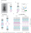
Figure 3. Super-cluster for centromere-kinetochore multimerisation
(a) left Cryo-electron microscopy image of isolated budding yeast kinetochore particles bound to microtubules. In the image, globular domains contact the microtubule which is encircled by a ring-like structure, likely DAM1C. There is also a central hub which does not contact the microtubule directly. Right Model for the architecture of a single k-unit where one kinetochore superassembly contacts one microtubule, as in budding yeast. Each CCAN anchors cohesin, which forms intramolecular loops on each side of the kinetochore. (b) Model for sister kinetochore coorientation during meiosis I in budding yeast. Two k-units - the sister kinetochores - are clamped together in a side-by-side orientation due to two kinds of linkages. (1) Monopolin binds to the Dsn1 subunit of the MIS12C and fuses the sister kinetochore together. (2) Meikin-Polo associates with CenpC/Mif2 and promotes coorientation, possibly by facilitating cohesin-dependent-linkages of sister centromeres. Note that a fused pair of sister kinetochores binds a single microtubule in meiosis I (261). (c) Schematic showing the architecture of the budding yeast kinetochore in mitosis in the presence and absence of spindle tension. Left: CCAN-anchored cohesin extrudes a loop on either side of the centromere until blocked by convergent genes at pericentromere borders. Borders also retain inter-sister, cohesive cohesin. This state, as shown, is short-lived because the attachment of sister kinetochores to microtubules from opposite poles results in the generation of tension. Right Under tension, the chromatin loops extend into a V-shaped structure. Intra-molecular, loop-extruding cohesin slides off but inter-molecular, cohesive, cohesin is trapped at the borders and holds the sister chromatids together. (d) Speculative model for the architecture of the mammalian kinetochore, inspired by the structure of the budding yeast kinetochore and pericentromeric chromatin in mitosis and meiosis. Ordered arrays of k-units are clustered together. This clustering is facilitated by cohesin anchored on CCAN and stabilised by cross-linkers between KMN, analogus to monopolin. Chromatin-organising complexes such as condensin may further serve to stabilize interactions between adjacent K-units.

Similar articles

Cited by

References

    1. Abad MA, Medina B, Santamaria A, Zou J, Plasberg-Hill C, et al. Structural basis for microtubule recognition by the human kinetochore Ska complex. Nat Commun. 2014;5 - PMC - PubMed
    1. Abad MA, Zou J, Medina-Pritchard B, Nigg EA, Rappsilber J, et al. Ska3 Ensures Timely Mitotic Progression by Interacting Directly With Microtubules and Ska1 Microtubule Binding Domain. Sci Reports. 2016;6(1):1–9. 2016 61. - PMC - PubMed
    1. Akiyoshi B, Nelson CR, Biggins S. The aurora B kinase promotes inner and outer kinetochore interactions in budding yeast. Genetics. 2013;194(3):785–89. - PMC - PubMed
    1. Allan LA, Camacho Reis M, Ciossani G, Huis in ‘t Veld PJ, Wohlgemuth S, et al. Cyclin B1 scaffolds MAD1 at the kinetochore corona to activate the mitotic checkpoint. EMBO J. 2020;39(12) - PMC - PubMed
    1. Allshire RC, Karpen GH. Epigenetic regulation of centromeric chromatin: old dogs, new tricks? Nat Rev Genet. 2008;9(12):923–37. - PMC - PubMed

Publication types

LinkOut - more resources