Review
. 2022 Sep;45(3):321-334.
doi: 10.1016/j.psc.2022.05.001. Epub 2022 Jul 31.

Neuropsychiatric Model of Addiction Simplified

Affiliations
Review

Neuropsychiatric Model of Addiction Simplified

Wilson M Compton et al. Psychiatr Clin North Am. 2022 Sep.

Abstract

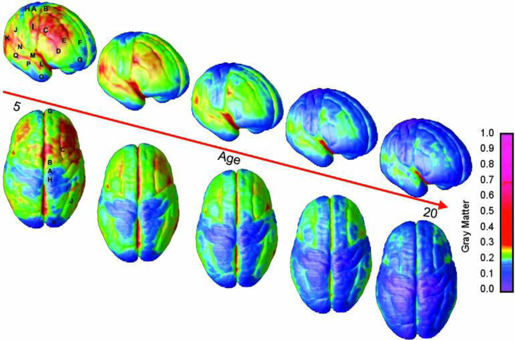
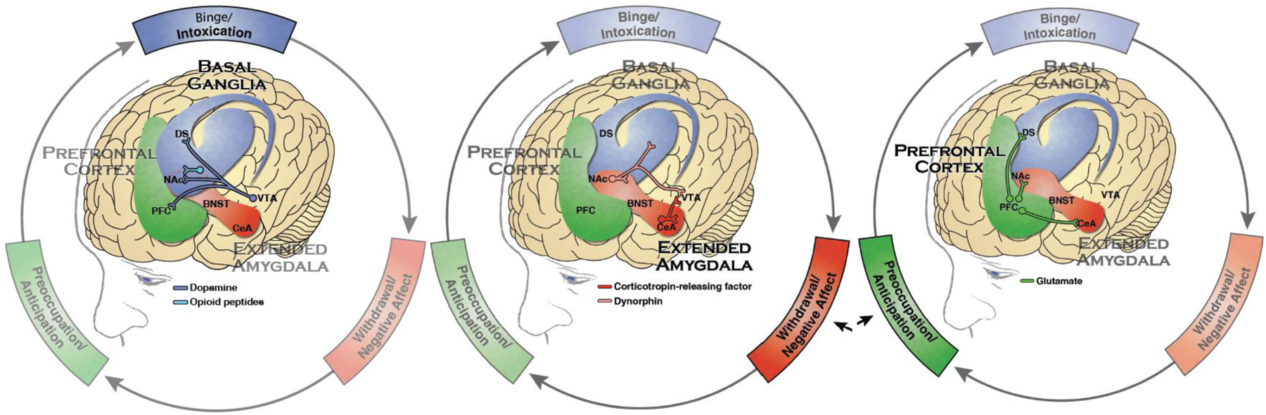
While substance experimentation typically begins in adolescence, substance use disorders (SUDs) usually develop in late teens or early adulthood, often in individuals who are vulnerable because of biological and socioeconomic risk factors. Severe SUDs-synonymous with addiction-involve changes in limbic and prefrontal brain areas after chronic drug exposure. These changes involve learned associations between drug reward and cues that trigger the anticipation of that reward (known as incentive salience), as well as heightened dysphoria during withdrawal and weakened prefrontal circuits needed for inhibiting habitual responses.

Keywords: Addiction; Addiction cycle; Neurobiology; Reinforcement.


Figures

Figure 1:
Figure 1:
Right lateral and top views of the dynamic sequence of gray matter maturation over the cortical surface. The side bar shows a color representation in units of gray matter volume. The following regions were selected for analyses in each hemisphere: A, precentral gyrus and primary motor cortex; B, superior frontal gyrus, posterior end near central sulcus; C, inferior frontal gyrus, posterior end; D, inferior frontal sulcus, anterior end in the ventrolateral prefrontal cortex; E, inferior frontal sulcus in the dorsolateral prefrontal cortex; F, anterior limit of superior frontal sulcus; G, frontal pole; H, primary sensory cortex in postcentral gyrus; I, supramarginal gyrus (area 40); J, angular gyrus (area 39); K, occipital pole; L–N, anterior, middle, and posterior portions of STG; O–Q, anterior, middle, and posterior points along the inferior temporal gyrus anterior end. (Reproduced from Gogtay N, Giedd JN, Lusk L, et al. Dynamic mapping of human cortical development during childhood through early adulthood. PNAS. 2004;101(21):8174–8179.)
Figure 2:
Figure 2:
The three stages of addiction: Binge/Intoxication, the stage at which an individual consumes an intoxicating substance and experiences its rewarding or pleasurable effects, primarily involves basal ganglia structures; Withdrawal/Negative Affect, the stage at which an individual experiences a negative emotional state in the absence of the substance, involves stress hormone responses and the extended amygdala; and Preoccupation/Anticipation, the stage at which one seeks substances again after a period of abstinence, involving interactions of the prefrontal cortex, the extended amygdala, and the basal ganglia. (Reproduced from U.S. Department of Health and Human Services (HHS), Office of the Surgeon General, Facing Addiction in America: The Surgeon General’s Report on Alcohol, Drugs, and Health. Washington, DC: HHS, 2016.) Notes: Blue represents the basal ganglia involved in the Binge/Intoxication stage. Red represents the extended amygdala involved in the Negative Affect/Withdrawal stage. Green represents the prefrontal cortex involved in the Preoccupation/Anticipation stage. Not shown is the neurotransmitter norepinephrine which is also activated in the extended amygdala during withdrawal. PFC - prefrontal cortex, DS - dorsal striatum, NAc - nucleus accumbens, BNST - bed nucleus of the stria terminalis, CeA - central nucleus of the amygdala, VTA - ventral tegmental area.

References

    1. O’Driscoll K, Leach JP. “No longer Gage”: an iron bar through the head. Early observations of personality change after injury to the prefrontal cortex. BMJ. 1998;317(7174):1673–1674. - PMC - PubMed
    1. American Psychiatric Association. Diagnostic and Statistical Manual of Mental Disorders, Fifth Edition, Text Revision (DSM-5-TR). Washington, D.C.: American Psychiatric Press. 2022.
    1. Koob GF, Volkow ND. Neurobiology of addiction: a neurocircuitry analysis. Lancet Psychiatry. 2016;3(8):760–773. - PMC - PubMed
    1. Koob GF, Volkow ND. Neurocircuitry of Addiction. Neuropsychopharmacology. 2010;35:217–238. - PMC - PubMed
    1. National Research Council and Institute of Medicine. From Neurons to Neighborhoods: The Science of Early Childhood Development. Committee on Integrating the Science of Early Childhood Development. Washington, D.C.: National Academy Press. 2000 - PubMed

Key Additional Readings:

    1. Volkow ND, Koob GF, McLellan AT. Neurobiologic Advances from the Brain Disease Model of Addiction. New England Journal of Medicine. 2016;374(4):363–371. - PMC - PubMed
    1. Compton WM, Valentino RJ, DuPont RL. Polysubstance use in the U.S. opioid crisis. Molecular Psychiatry. 2021;26(1):41–50. - PMC - PubMed
    1. Volkow ND, Boyle M. Neuroscience of addiction: relevance to prevention and treatment. American Journal of Psychiatry. 2018;175:729–740. - PubMed
    1. Feltenstein MW, See RE, Fuchs RA. Neural Substrates and Circuits of Drug Addiction. Cold Spring Harbor Perspectives in Medicine. 2021;11(4):a039628. - PMC - PubMed