. 2022 Sep 2;13(9):760.
doi: 10.1038/s41419-022-05207-8.

Nintedanib induces senolytic effect via STAT3 inhibition

Affiliations

Nintedanib induces senolytic effect via STAT3 inhibition

Hyun-Ji Cho et al. Cell Death Dis. .

Abstract

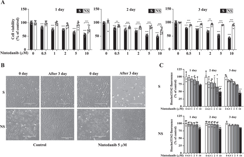
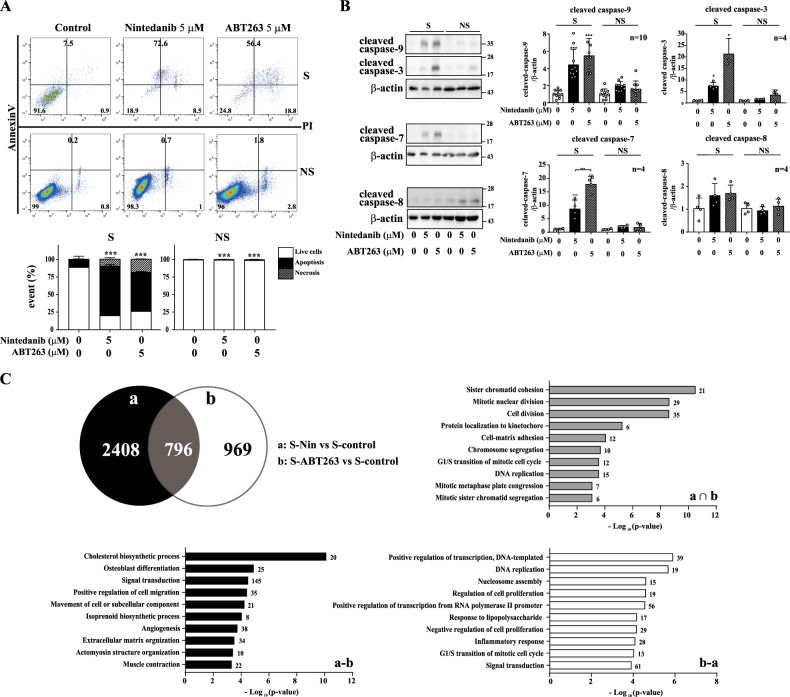
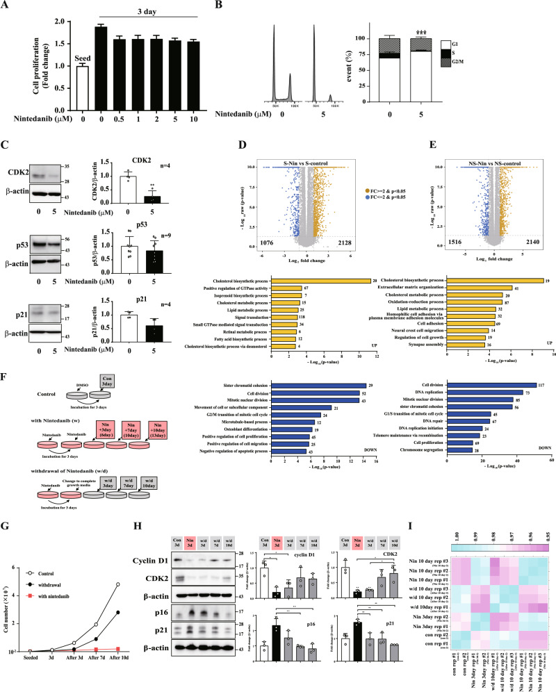
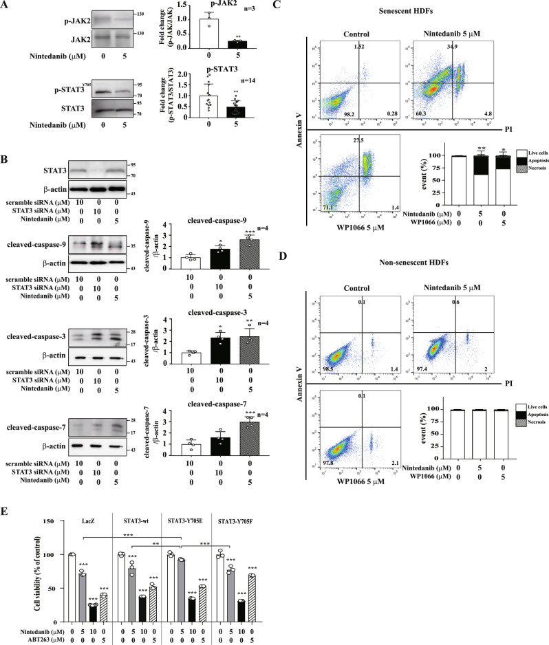
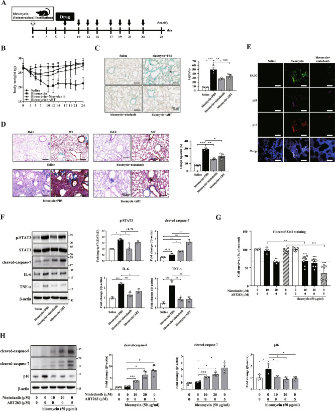
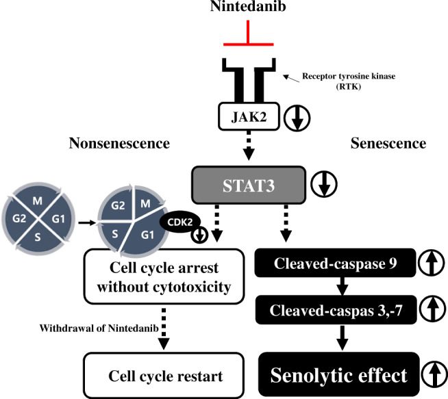
Selective removal of senescent cells, or senolytic therapy, has been proposed to be a potent strategy for overcoming age-related diseases and even for reversing aging. We found that nintedanib, a tyrosine kinase inhibitor, selectively induced the death of primary human dermal fibroblasts undergoing RS. Similar to ABT263, a well-known senolytic agent, nintedanib triggered intrinsic apoptosis in senescent cells. Additionally, at the concentration producing the senolytic effect, nintedanib arrested the cell cycle of nonsenescent cells in the G1 phase without inducing cytotoxicity. Interestingly, the mechanism by which nintedanib activated caspase-9 in the intrinsic apoptotic pathway differed from that of ABT263 apoptosis induction; specifically, nintedanib did not decrease the levels of Bcl-2 family proteins in senescent cells. Moreover, nintedanib suppressed the activation of the JAK2/STAT3 pathway, which caused the drug-induced death of senescent cells. STAT3 knockdown in senescent cells induced caspase activation. Moreover, nintedanib reduced the number of senescence-associated β-galactosidase-positive senescent cells in parallel with a reduction in STAT3 phosphorylation and ameliorated collagen deposition in a mouse model of bleomycin-induced lung fibrosis. Consistently, nintedanib exhibited a senolytic effect through bleomycin-induced senescence of human pulmonary fibroblasts. Overall, we found that nintedanib can be used as a new senolytic agent and that inhibiting STAT3 may be an approach for inducing the selective death of senescent cells. Our findings pave the way for expanding the senolytic toolkit for use in various aging statuses and age-related diseases.


Conflict of interest statement

The authors declare no competing interests.

Figures

Fig. 1
Fig. 1. Nintedanib reduces the viability of senescent HDFs.
A Senescent (S) HDFs and nonsenescent (NS) HDFs were treated with nintedanib (0.5, 1, 2, 5, and 10 μM) for 1, 2, or 3 days, and then CCK-8 assays were performed to assess cell viability. n = 8. B Cell morphological changes after three days of treatment with DMSO (control) or nintedanib (5 μM). Senescent (S) HDFs (5 × 104 cells per well) and nonsenescent (NS) HDFs (8 × 104 cells per well) were separately plated in 6-well plates. Images were randomly captured by inverted microscopy. Scale bar, 500 μm. C Senescent (S) HDFs and nonsenescent (NS) HDFs were treated with DMSO or nintedanib (0.5, 1, 2, 5, and 10 μM) for three days, and Hoechst 33342 staining was then performed to investigate cell growth. The data normalized to those for DMSO-treated cells are shown as the mean ± S.D. *p < 0.05, **p < 0.01, ***p < 0.001 by one-way ANOVA with Tukey’s post hoc test.
Fig. 2
Fig. 2. Nintedanib induces apoptotic cell death in senescent HDFs.
A Senescent (S) and nonsenescent (NS) HDFs were treated with DMSO, nintedanib (5 μM), or ABT263 (5 μM) for three days, and flow cytometry analysis was performed using Annexin V/PI staining to identify apoptotic cells. n = 4. B Senescent (S) and nonsenescent (NS) HDFs were treated with DMSO, nintedanib (5 μM), or ABT263 (5 μM) for three days, and then western blot assays using anti-caspase-9, anti-caspase-3, anti-caspase-7, and anti-caspase-8 antibodies were performed to investigate the apoptotic pathways involved in the senolytic effect. n = 4–10. C Venn diagram (top left) showing the number of differentially expressed genes (DEGs) in senescent cells treated with nintedanib (Group a) or treated with ABT263 (Group b). A Gene Ontology biological process (GOBP) analysis (top right, bottom left and bottom right) of each set (a∩b, a-b, and b-a, respectively). The data normalized to those for DMSO-treated cells are shown as the mean ± S.D. *p < 0.05, **p < 0.01, ***p < 0.001 by one-way ANOVA with Tukey’s post hoc test.
Fig. 3
Fig. 3. Nintedanib induces G1-phase cell cycle arrest without inducing cell death in nonsenescent HDFs.
A Nonsenescent (NS) HDFs were treated with nintedanib (0.5, 1, 2, 5, and 10 μM) for 3 days, and then CCK-8 assays were performed to evaluate cell viability and proliferation. n = 8. B Nonsenescent (NS) HDFs were treated with DMSO or nintedanib (5 μM) for one day and flow cytometry analysis of PI staining was performed to determine the number of cells in each cell cycle phase. n = 5. C Nonsenescent (NS) HDFs were treated with DMSO or nintedanib (5 μM) for one day, and then western blot assays using anti-CDK2, anti-p53, and anti-p21 antibodies were performed to investigate the protein expression of cell cycle regulators. n = 4–9. D Volcano plot (top) and GOBP analysis (bottom) of up- (yellow) and downregulated (blue) DEGs in nintedanib-treated senescent cells. E Volcano plot (top) and GOBP analysis (bottom) of up- (yellow) and downregulated (blue) DEGs in nintedanib-treated nonsenescent cells. F Experimental scheme for testing the restoration of cell proliferation after nintedanib removal. The ‘control’ group was treated with DMSO and the ‘Nin n day’ group was treated with nintedanib for n days. The ‘withdrawal’ group was treated with nintedanib for 3 days and then cultured in complete growth medium from 3 to 10 days after drug removal. G Nonsenescent HDFs were treated according to the experimental scheme, and the number of cells was monitored to investigate the effect of drug removal on cell proliferation. H Nonsenescent HDFs were treated according to the experimental scheme, and then western blot assays using anti-cyclin D1, anti-CDK2, anti-p16, and anti-p21 antibodies were performed to investigate the restoration of cell proliferation after drug removal. n = 3. I Correlation matrix for the transcriptome of all samples shown in G. The data normalized to those for DMSO-treated cells are shown as the mean ± S.D. *p < 0.05, **p < 0.01, ***p < 0.001 by one-way ANOVA with Tukey’s post hoc test.
Fig. 4
Fig. 4. Nintedanib inhibits tyrosine phosphorylation of JAK2 and STAT3 in senescent HDFs.
A Senescent (S) HDFs were treated with DMSO or nintedanib (5 μM) for one hour, and then western blot assays using anti-p-JAK2, anti-JAK2, anti-p-STAT3, and anti-STAT3 antibodies were performed to identify the signaling pathways affected by nintedanib. B Senescent (S) HDFs were transfected with scramble siRNA or STAT3-specific siRNA three days prior to nintedanib (5 μM) treatment. Then, after drug exposure for three days, western blot assays using anti-STAT3, anti-caspase-9, anti-caspase-3, and anti-caspase-7 antibodies were performed to evaluate apoptosis induction. C Senescent (S) HDFs were treated with DMSO, nintedanib (5 μM), or WP1066 (5 μM, a STAT3 kinase inhibitor) for three days, and a flow cytometry analysis with Annexin V/PI staining was performed to identify apoptotic cells. D Nonsenescent (NS) HDFs were treated with DMSO, nintedanib (5 μM), or WP1066 (5 μM, STAT3 kinase inhibitor) for three days, and flow cytometry analysis with Annexin V/PI staining was performed to identify apoptotic cells. E Nonsenescent HDFs were infected with lentiviral particles having LacZ (as control), STAT3-wt, STAT3-Y705E, or STAT3-Y705F encoded gene and then CCK-8 assays were performed after nintedanib treatment for 3 days to assess cell viability. n = 3. The data normalized to those for DMSO-treated cells are shown as the mean ± S.D. *p < 0.05, **p < 0.01, ***p < 0.001 by one-way ANOVA with Tukey’s post hoc test.
Fig. 5
Fig. 5. Nintedanib shows senolysis in bleomycin-induced in vivo and in vitro model.
A Experimental scheme for establishing bleomycin-induced lung fibrosis model: saline (n = 3), PBS (bleomycin; n = 2), ABT263 (bleomycin + ABT; n = 3), or nintedanib (bleomycin + nintedanib; n = 4) was administrated via intraperitoneal injection three times per week for 24 days (black arrow). Control mice were treated with PBS are labeled saline (n = 3). B Body weight changes after bleomycin followed by drug treatment. C, D Lung tissues were stained with an SAβG staining kit to measure the number of senescent cells (C), hematoxylin and eosin (H&E) to assess lung interstitial damage, and Masson’s trichrome (MT) staining (collagen is stained blue) to detect collagen deposition (D). Scale bar, 200 μm. E Lung tissues were stained with SPiDER-βGal to measure SAβG activity and with anti-p16 and anti-p53 antibodies to determine whether senescence was attenuated. Scale bar, 200 μm. F Western blot assays using anti-p-STAT3, anti-STAT3, anti-caspase-7, anti-IL-6 and anti-TNF-α antibodies were performed to identify the apoptotic pathways involved in the lung tissue. G Bleomycin-induced senescent HPFs was treated with nintedanib or ABT263 for 3 days. Then, Hoechst 33342 staining was performed to assess cell viability. n = 3 H Bleomycin-induced senescent HPFs were treated with nintedanib or ABT263 for 3 days, and then, western blot assays using anti-caspase-9, anti-caspase-7, and anti-p16 antibodies were performed to identify the apoptotic pathways involved in the senolytic effect. The data normalized to those for DMSO-treated cells are shown as the mean ± S.D (F, G). *p < 0.05, **p < 0.01, ***p < 0.001 by one-way ANOVA with Tukey’s post hoc test.
Fig. 6
Fig. 6. Nintedanib induces a senolytic effect in senescent HDFs via JAK2/STAT3 pathway.
Proposed mechanism of senolytic effect induced by nintedanib. Nintedanib-induced senescent cell death is mediated by inhibiting STAT3 activity. Additionally, nintedanib causes cell cycle arrest in nonsenescent cells by regulating CDK2.

References

    1. Papadopoulos D, Magliozzi R, Mitsikostas DD, Gorgoulis VG, Nicholas RS. Aging, cellular senescence, and progressive multiple sclerosis. Front Cell Neurosci. 2020;14:178. doi: 10.3389/fncel.2020.00178. - DOI - PMC - PubMed
    1. Di Micco R, Krizhanovsky V, Baker D, d’Adda, di Fagagna F. Cellular senescence in ageing: from mechanisms to therapeutic opportunities. Nat Rev Mol Cell Biol. 2021;22:75–95. doi: 10.1038/s41580-020-00314-w. - DOI - PMC - PubMed
    1. Zhavoronkov A, Bhullar B. Classifying aging as a disease in the context of ICD-11. Front Genet. 2015;6:326. doi: 10.3389/fgene.2015.00326. - DOI - PMC - PubMed
    1. Khaltourina D, Matveyev Y, Alekseev A, Cortese F, Iovita A. Aging fits the disease criteria of the international classification of diseases. Mech Ageing Dev. 2020;189:111230. doi: 10.1016/j.mad.2020.111230. - DOI - PubMed
    1. Niedernhofer LJ, Robbins PD. Senotherapeutics for healthy ageing. Nat Rev Drug Disco. 2018;17:377. doi: 10.1038/nrd.2018.44. - DOI - PubMed

Publication types