. 2022 Sep 2;12(1):14971.
doi: 10.1038/s41598-022-18977-0.

Low quality antibody responses in critically ill patients hospitalized with pandemic influenza A(H1N1)pdm09 virus infection

Affiliations

Low quality antibody responses in critically ill patients hospitalized with pandemic influenza A(H1N1)pdm09 virus infection

Xiuhua Lu et al. Sci Rep. .

Abstract

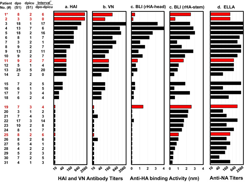
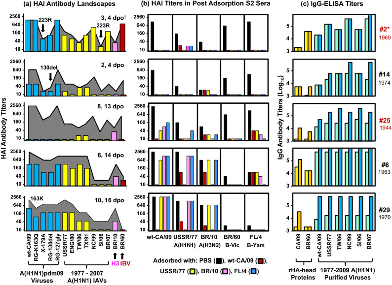
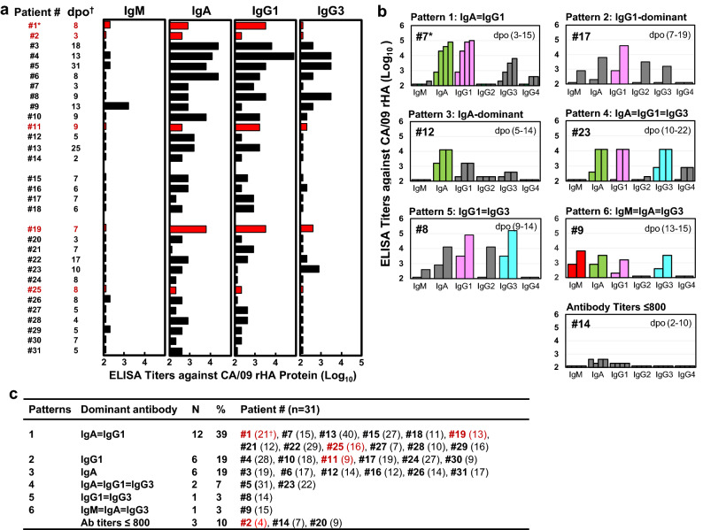
Although some adults infected with influenza 2009 A(H1N1)pdm09 viruses mounted high hemagglutination inhibition (HAI) antibody response, they still suffered from severe disease, or even death. Here, we analyzed antibody profiles in patients (n = 31, 17-65 years) admitted to intensive care units (ICUs) with lung failure and invasive mechanical ventilation use due to infection with A(H1N1)pdm09 viruses during 2009-2011. We performed a comprehensive analysis of the quality and quantity of antibody responses using HAI, virus neutralization, biolayer interferometry, enzyme-linked-lectin and enzyme-linked immunosorbent assays. At time of the ICU admission, 45% (14/31) of the patients had HAI antibody titers ≥ 80 in the first serum (S1), most (13/14) exhibited narrowly-focused HAI and/or anti-HA-head binding antibodies targeting single epitopes in or around the receptor binding site. In contrast, 42% (13/31) of the patients with HAI titers ≤ 10 in S1 had non-neutralizing anti-HA-stem antibodies against A(H1N1)pdm09 viruses. Only 19% (6/31) of the patients showed HA-specific IgG1-dominant antibody responses. Three of 5 fatal patients possessed highly focused cross-type HAI antibodies targeting the (K130 + Q223)-epitopes with extremely low avidity. Our findings suggest that narrowly-focused low-quality antibody responses targeting specific HA-epitopes may have contributed to severe infection of the lower respiratory tract.


Conflict of interest statement

The authors declare no competing interests.

Figures

Figure 1
Figure 1
A(H1N1)pdm09 virus infections induced low quality antibody responses in most patients at the time of ICU admission. The first serum samples (S1) collected 1 to 7 days post ICU admission (dpicu) and 2 to 25 days post-symptom onset (dpo) from 31 patients were tested by: (a) HAI assays using wt-CA/09 (Q223), (b) VN assays using MX/09 (Q223QR); (c) BLI assays using rHA-head from wt-CA/09 and rHA-stem from A/Michigan/45/2015, and (d) ELLA assays using A(H6N1) reassortant virus possessing wt-CA/09 NA. Antibody responses are illustrated by black bars for survivors and red bars for patients with fatal outcomes. For each sample, we completed three independent HAI and VN assays. ELLA assays were performed in duplicate. BLI assays were performed in 2 independent assays. *The interval between S1 dpo and S1 dpicu, and fatal patients in red.
Figure 2
Figure 2
Kinetics of anti-HA antibody responses. Serum samples were tested by HAI assays using wt-CA/09, VN assays using MX/09, and BLI assays using rHA-head from wt-CA/09 and rHA-stem from A/Michigan/45/2015. (a) HAI and VN antibody response kinetics in 8 patients are illustrated by black lines and red lines, respectively. (b) Anti-HA-head and anti-HA-stem antibody response kinetics are illustrated by black lines and red lines, respectively. (c) Ratio of anti-head/stem ABA in 31 patients was categorized into 5 age-groups. Each colored square in 5 age-groups represents the ratio in each serum sample. Each color(yellow, orange, blue, dark blue, green, cyan, brown, red, and pink) in each age group represented the ratio(s) from the same patient’s serum sample(s) that were collected 1–4 times at 3 different time periods of 2–7 dpo, 8–14 dpo, and 15–45 dpo. Red and pink square with patient numbers represented 5 fatal patients; other color squares without patient numbers represented 26 surviving patients. (d) Summary of the percentage of 31 patients with ratio of anti-head/stem ABA < 0.6. We completed 3 independent HAI and VN assays. BLI assays were performed in 2 independent assays.
Figure 3
Figure 3
Determination of immunodominance of anti-HA-head binding antibody responses and epitope mapping. Anti-HA-head ABAs were determined by BLI assays using a rHA1-wt (HA-head from CA/09) and 15 rHA1-mutants possessing single point mutations or a K130 deletion (130del). (a) The 12 patients displayed focused anti-head ABA [defined by > 50% ABA reduction against rHA1-mutant(s) compared to rHA1-wt]. Patients #7 and #12 showing completely focused ABA are highlighted in blue. (b) Summary of key contact aa determined by HAI assays and BLI assays. Key contact aa are determined as virus-mutants or rHA1-mutants causing ≥ fourfold HAI antibody reduction in HAI assays or > 50% ABA reduction in BLI assays, respectively. (c) Key contact residues are mapped onto the CA/09 HA-head structure monomer. Antigenic sites Sa (red), Sb (Magenta), Ca (Blue), Receptor binding site (RBS, orange). *Serum collection days post-symptom onset (dpo) and post ICU admission (dpicu). R, RBS. ND, not determined.
Figure 4
Figure 4
HAI antibodies targeting epitopes possessing HA-(K130 + Q223) cross-reacted with A(H3N2) IAV or IBVs. (a) HAI antibody landscapes in 5 patients were constructed using HAI assays with the 14 indicated viruses. HAI titers in S1 were shown in different colored bars: blue bars for wt-CA/09 and CA/09-mutants, yellow bars for 1977–2007 A(H1N1), pink bars for BR/10 A(H3N2), and red bars for BR/60 IBV. HAI antibody titers in the second sera are shown in gray landscapes. (b) Sera were adsorbed with purified viruses or PBS as a control. Post adsorption sera were tested by HAI assays with wt-CA/09, USSR/77, BR/10, BR/60 and FL/4. Antibody titers are expressed as color bars: post-adsorbed with PBS in black, wt-CA/09 in red, USSR/77 in yellow, BR/10 in pink, and FL/4 in blue. We completed two independent antibody adsorption assays. (c) Paired sera were tested by IgG-ELISA using two rHA-heads from CA/09 or BR/60 and 6 purified A(H1N1) viruses. IgG titers are shown in yellow or light blue bars for S1 and orange or dark blue bars for the second sera. We completed two independent ELISA assays. *Patient number (fatal patients in red) with birth year. Serum collection days post-symptom onset (dpo).
Figure 5
Figure 5
CA/09 HA-specific antibody isotype and IgG subclass responses. Sera were tested by ELISA using rHA from wt-CA/09. (a) IgM, IgA, IgG1, and IgG3 antibody responses in 31 patients. ELISA antibody titers are illustrated by black bars for survivors and red bars for fatal patients. (b) Representative patients showed 6 different antibody isotype and IgG subclass immunodominance response patterns. Antibody titers are shown in gray bars, but dominant and co-dominant isotype and IgG subclass are highlighted in other colors: IgG1 in pink, IgG3 in cyan, IgA in green, and IgM in red. (c) Summary of antibody isotype and IgG subclass response patterns at indicated serum collection time in 31 patients (Supplementary Table 4). *Fatal patients in red, serum collection days post-symptom onset (dpo). For each serum sample, we completed two independent ELISA assays.

References

    1. Paules C, Subbarao K. Influenza. Lancet. 2017;390:697–708. doi: 10.1016/S0140-6736(17)30129-0. - DOI - PubMed
    1. Katz JM, Hancock K, Xu X. Serologic assays for influenza surveillance, diagnosis and vaccine evaluation. Expert Rev. Anti Infect. Ther. 2011;9:669–683. doi: 10.1586/eri.11.51. - DOI - PubMed
    1. Dawood FS, et al. Estimated global mortality associated with the first 12 months of 2009 pandemic influenza A H1N1 virus circulation: A modelling study. Lancet Infect. Dis. 2012;12:687–695. doi: 10.1016/S1473-3099(12)70121-4. - DOI - PubMed
    1. Krammer F. The human antibody response to influenza A virus infection and vaccination. Nat. Rev. Immunol. 2019;19:383–397. doi: 10.1038/s41577-019-0143-6. - DOI - PubMed
    1. Boudreau CM, Alter G. Extra-neutralizing FcR-mediated antibody functions for a universal influenza vaccine. Front. Immunol. 2019;10:440. doi: 10.3389/fimmu.2019.00440. - DOI - PMC - PubMed