

Neuroimaging in schizophrenia: an overview of findings and their implications for synaptic changes
- PMID: 36056106
- PMCID: PMC9700830
- DOI: 10.1038/s41386-022-01426-x
Neuroimaging in schizophrenia: an overview of findings and their implications for synaptic changes
Abstract
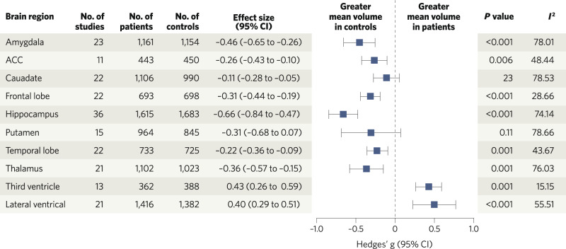
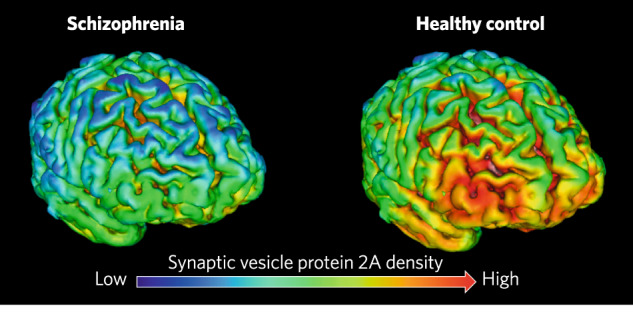
Over the last five decades, a large body of evidence has accrued for structural and metabolic brain alterations in schizophrenia. Here we provide an overview of these findings, focusing on measures that have traditionally been thought to reflect synaptic spine density or synaptic activity and that are relevant for understanding if there is lower synaptic density in the disorder. We conducted literature searches to identify meta-analyses or other relevant studies in patients with chronic or first-episode schizophrenia, or in people at high genetic or clinical risk for psychosis. We identified 18 meta-analyses including over 50,000 subjects in total, covering: structural MRI measures of gyrification index, grey matter volume, grey matter density and cortical thickness, neurite orientation dispersion and density imaging, PET imaging of regional glucose metabolism and magnetic resonance spectroscopy measures of N-acetylaspartate. We also review preclinical evidence on the relationship between ex vivo synaptic measures and structural MRI imaging, and PET imaging of synaptic protein 2A (SV2A). These studies show that schizophrenia is associated with lower grey matter volumes and cortical thickness, accelerated grey matter loss over time, abnormal gyrification patterns, and lower regional SV2A levels and metabolic markers in comparison to controls (effect sizes from ~ -0.11 to -1.0). Key regions affected include frontal, anterior cingulate and temporal cortices and the hippocampi. We identify several limitations for the interpretation of these findings in terms of understanding synaptic alterations. Nevertheless, taken with post-mortem findings, they suggest that schizophrenia is associated with lower synaptic density in some brain regions. However, there are several gaps in evidence, in particular whether SV2A findings generalise to other cohorts.
© 2022. The Author(s).
Conflict of interest statement
OH is a part-time employee of H Lundbeck A/s. He has received investigator-initiated research funding from and/or participated in advisory/speaker meetings organised by Angellini, Autifony, Biogen, Boehringer-Ingelheim, Eli Lilly, Heptares, Global Medical Education, Invicro, Jansenn, Lundbeck, Neurocrine, Otsuka, Sunovion, Recordati, Roche and Viatris/Mylan. Neither Dr Howes or his family have holdings/a financial stake in any pharmaceutical company. Dr Howes has a patent for the use of dopaminergic imaging. ES, GC and CC have reported no biomedical financial interests or potential conflicts of interest. The views expressed are those of the authors and not necessarily those of H Lundbeck A/s, the NHS/NIHR or the Department of Health.
Figures
References
-
- McCutcheon RA, Marques TR, Howes OD. Schizophrenia—an overview. JAMA Psychiatry. 2020;77:201–10. - PubMed
-
- Feinberg I. Schizophrenia: caused by a fault in programmed synaptic elimination during adolescence? J Psychiatr Res. 1982;17:319–34. - PubMed
-
- Keshavan MS, Anderson S, Pettergrew JW. Is schizophrenia due to excessive synaptic pruning in the prefrontal cortex? The Feinberg hypothesis revisited. J Psychiatr Res. 1994;28:239–65. - PubMed
-
- Howes OD, Shatalina E. Integrating the neurodevelopmental and dopamine hypotheses of schizophrenia and the role of cortical excitation-inhibition balance. Biol Psychiatry. 2022;92:501–13. - PubMed
Publication types
MeSH terms
Grants and funding
LinkOut - more resources
Full Text Sources
Medical
