. 2022 Sep 2;5(1):902.
doi: 10.1038/s42003-022-03875-y.

Spike-based adenovirus vectored COVID-19 vaccine does not aggravate heart damage after ischemic injury in mice

Affiliations

Spike-based adenovirus vectored COVID-19 vaccine does not aggravate heart damage after ischemic injury in mice

Shanshan Gu et al. Commun Biol. .

Abstract

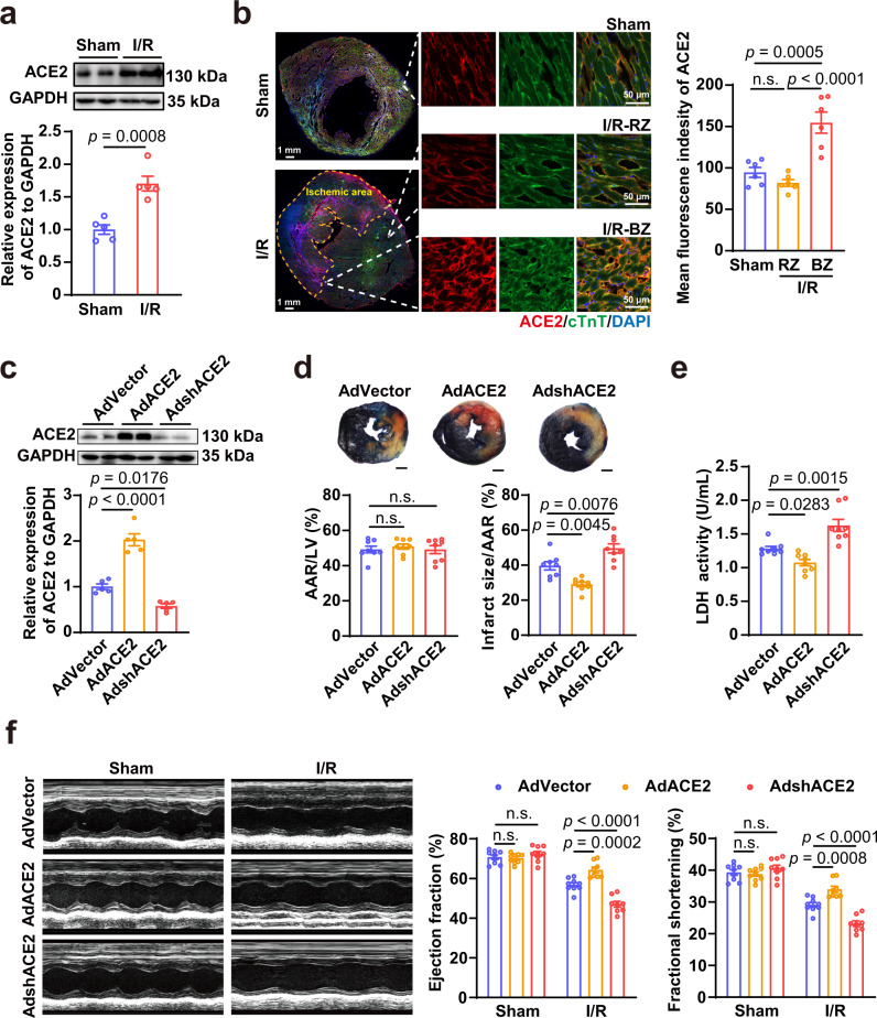
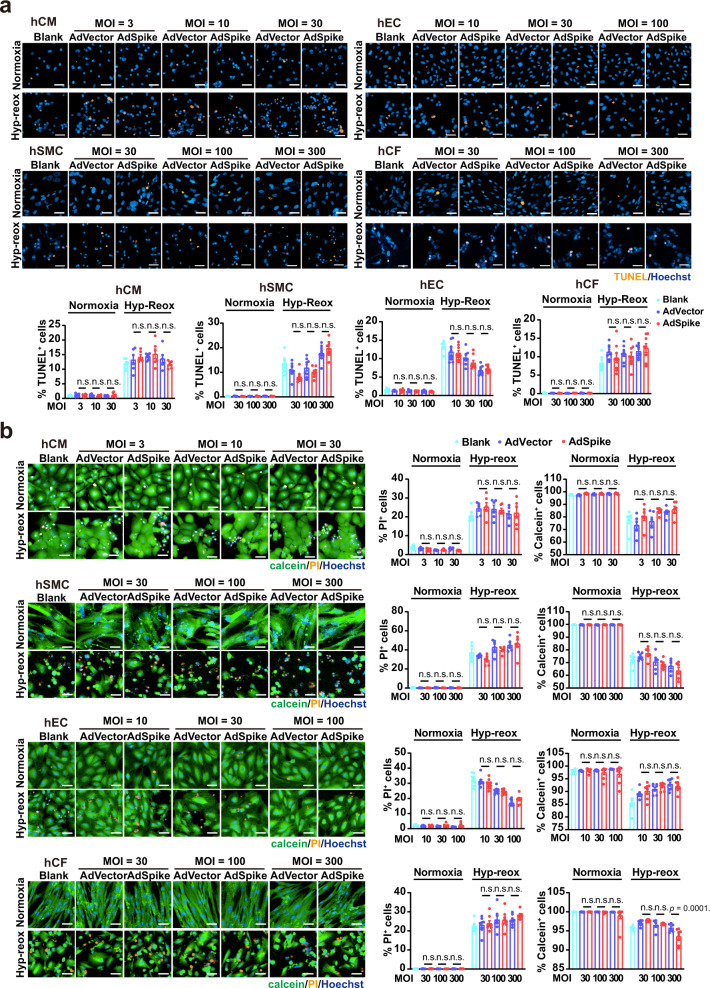
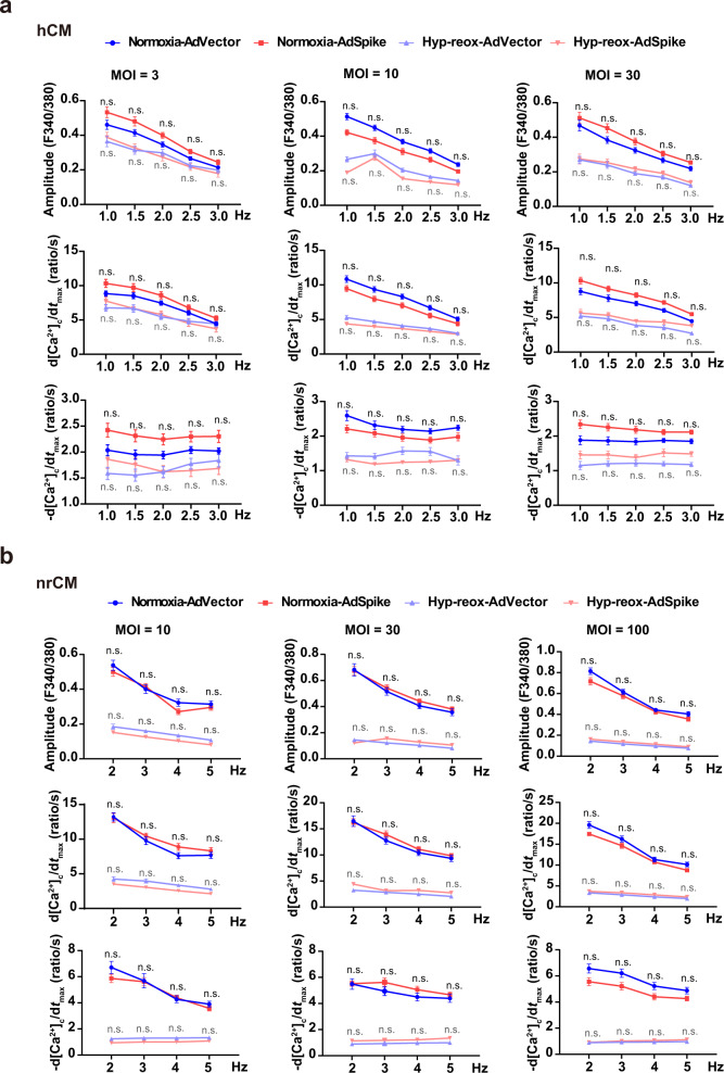
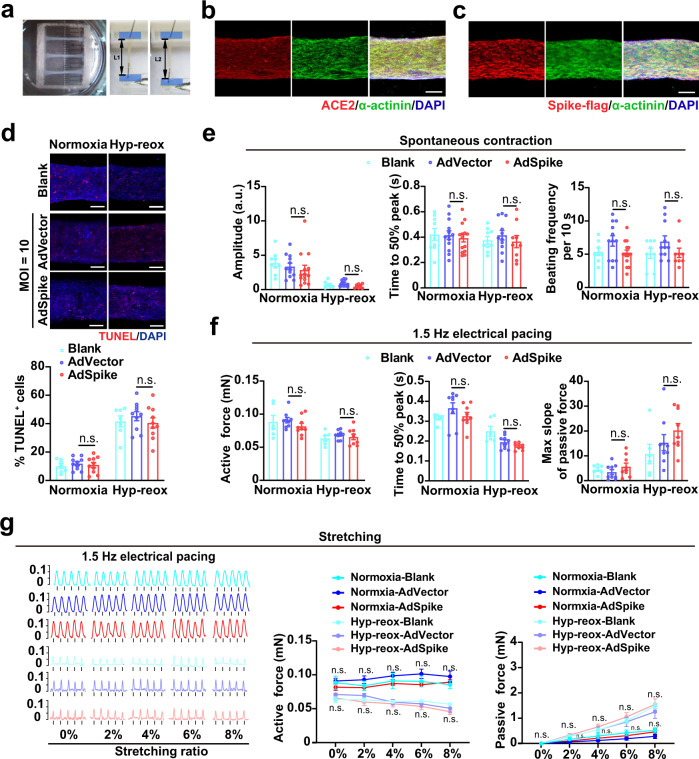
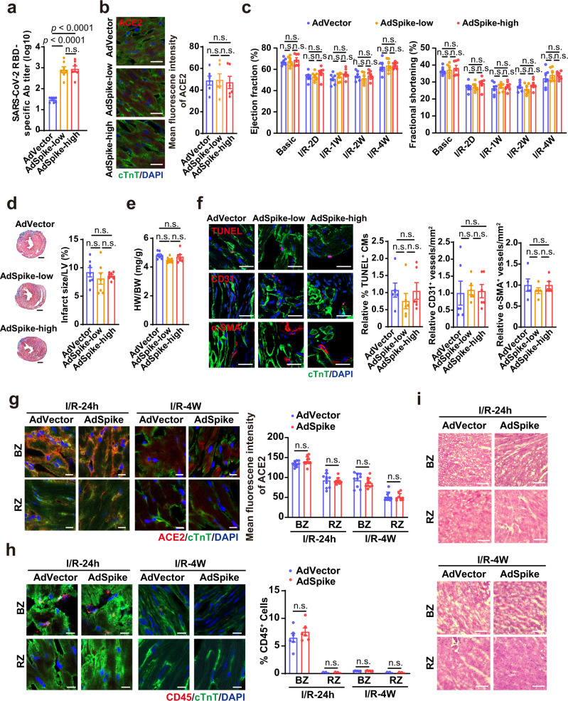
An unprecedented number of COVID-19 vaccination campaign are under way worldwide. The spike protein of SARS-CoV-2, which majorly binds to the host receptor angiotensin converting enzyme 2 (ACE2) for cell entry, is used by most of the vaccine as antigen. ACE2 is highly expressed in the heart and has been reported to be protective in multiple organs. Interaction of spike with ACE2 is known to reduce ACE2 expression and affect ACE2-mediated signal transduction. However, whether a spike-encoding vaccine will aggravate myocardial damage after a heart attack via affecting ACE2 remains unclear. Here, we demonstrate that cardiac ACE2 is up-regulated and protective after myocardial ischemia/reperfusion (I/R). Infecting human cardiac cells or engineered heart tissues with a spike-based adenovirus type-5 vectored COVID-19 vaccine (AdSpike) does not affect their survival and function, whether subjected to hypoxia-reoxygenation injury or not. Furthermore, AdSpike vaccination does not aggravate heart damage in wild-type or humanized ACE2 mice after I/R injury, even at a dose that is ten-fold higher as used in human. This study represents the first systematic evaluation of the safety of a leading COVID-19 vaccine under a disease context and may provide important information to ensure maximal protection from COVID-19 in patients with or at risk of heart diseases.


Conflict of interest statement

The authors declare no competing interests.

Figures

Fig. 1
Fig. 1. The protective effect of ACE2 during myocardial I/R injury.
a Representative and quantitative immunoblot analysis of ACE2 protein in the hearts after myocardial I/R (45 min/24 h). n = 5 for each group. b Representative immunostaining analysis of ACE2 expression with quantification of the mean fluorescence intensity in the hearts after myocardial I/R (45 min/24 h). RZ, remote zone; BZ, border zone. n = 6 for each group. c Representative and quantitative immunoblot analysis of ACE2 protein in the hearts infected with adenovirus-mediated vector control (AdVector), adenovirus-mediated ACE2-overexpression (AdACE2), or adenovirus-mediated ACE2 short hairpin RNA (AdshACE2) for 3 days. n = 5 for each group. d Representative 2, 3, 5-triphenyltetrazolium chloride (TTC)/Evans blue staining (upper) and averaged infarct size (lower) following myocardial I/R (45 min/24 h). AAR, area at risk; LV, left ventricle; n = 8 for each group; Scale bar, 1 mm. e The lactic dehydrogenase (LDH) activity in the serum of mice infected with AdVector, AdACE2, or AdshACE2 following myocardial I/R (45 min/24 h). n = 8 for each group. f Representative M-mode echocardiogram images and statistical analysis of ejection fraction and fractional shortening in the hearts infected with AdVector, AdACE2, or AdshACE2 following myocardial I/R (45 min/24 h). n = 8–9 for each group. Statistical significance was assessed using unpaired, two-tailed Student t test (a), one-way ANOVA with Turkey post-test (be) or 2-way ANOVA with Turkey post-test (f). Data are presented as mean ± SEM; n.s., not significant.
Fig. 2
Fig. 2. AdSpike has little effect on cell survival of human cardiac cells with or without hyp-reox injury.
a Representative and quantitative immunostaining analysis of the TUNEL+ cells in hCMs, hSMCs, hECs, and hCFs infected by AdVector or AdSpike with or without hyp-reox injury. Scale bar, 50 μm. n = 5–8 for each group. b Representative and quantitative calcein-AM/propidium iodide (PI) double staining in hCMs, hSMCs, hECs, hCFs infected by AdVector or AdSpike with or without hyp-reox injury. n = 5–8 for each group. Statistical significance was assessed using 2-way ANOVA with Turkey post-test. Scale bar, 50 μm. Data are presented as mean ± SEM; n.s., not significant.
Fig. 3
Fig. 3. AdSpike has little effect on calcium handling properties of cultured cardiomyocytes subjecting to electrical field stimulation with or without hyp-reox injury.
Averaged parameters of calcium transient of hCMs (a) or neonatal rat cardiomyocytes (nrCM, b) infected by AdVector or AdSpike with different frequency of electrical field stimulation. n = 15–30 for each group. Statistical significance was assessed using 2-way ANOVA with Turkey post-test. Data are presented as mean ± SEM; n.s., not significant as compared with the corresponding AdVector control, in which the black color indicates comparison within the normoxia group, and gray color indicates comparison within the hyp-reox group.
Fig. 4
Fig. 4. AdSpike has little effect on hEHTs that underwent spontaneous contraction, electrical field stimulation, and mechanical tensile test with or without hyp-reox injury.
a Low magnification image of hEHT on day 21 (left) and the equipment image of mechanical contractility force test for hEHT (right). Scale bar, 2 mm. b, c Co-localization analysis of anti-α-actinin (green) and anti-ACE2 (red) or anti-Spike-flag (red) with DAPI (blue). Scale bar, 200 μm. d Representative image and quantification of TUNEL staining in hEHTs with or without hyp-reox injury. n = 7–10 for each group. Scale bar, 200 μm. e Video analysis of the hEHTs for spontaneous contraction amplitude, time to 50% peak, and beating frequency per 10 seconds. a.u., absolute units. n = 9–14 for each group. f The active contractile force, time to 50% peak and maximum slope of passive force of the bundles at 1.5 Hz electrical pacing with or without hyp-reox injury. n = 6–9 for each group. g Representative contractile force traces and active contractile force during progressive stretching (0%, 2%, 4%, 6%, and 8% tissue length) of hEHTs with or without hyp-reox injury on day 22. n = 5–9 for each group. Statistical significance was assessed using 2-way ANOVA with Turkey post-test. Data are presented as mean ± SEM; n.s., not significant, compared with the corresponding AdVector control, in which the black color indicates comparison within the normoxia group, and gray color indicates comparison within the hyp-reox group.
Fig. 5
Fig. 5. AdSpike has little effect on heart damage repair after myocardial I/R on hACE2 mice in vivo.
a Antibody tilters of SARS-CoV-2 spike receptor-binding domain (RBD) in the mouse serum collected at 4 weeks after injection of AdSpike at various doses. n = 7 for each group. b Representative immunostaining analysis of the ACE2+ cardiomyocytes with quantification of the mean fluorescence intensity in the heart at 4 weeks post-vaccination. n = 6 for each group. Scale bar, 20 μm. c Ejection fraction and fractional shortening of hACE2 mice heart after I/R measured by echocardiography at various time points post-I/R measured by echocardiography. D, day; W, week. n = 7 for each group. d Masson-Trichrome staining of heart cross sections of hACE2 mice 4 weeks post-I/R (left) with quantification of scar size (right). n = 7 for each group. Scale bar, 1 mm. e Quantification of heart weight (HW) relative to body weight (BW) of hACE2 mice at 4 weeks post-I/R. n = 7 for each group. f Representative and quantitative immunostaining analysis of TUNEL+/cTnT+ cardiomyocytes, CD31+ capillaries, and α-SMA+ vessels in the border zones of infarcted hACE2 mouse hearts 4 weeks post-I/R. n = 5–6 for each group. Scale bar, 50 μm g Representative immunostaining analysis of the ACE2+ cardiomyocytes with quantification of the mean fluorescence intensity in border zone (BZ) or remote zone (RZ) at 24 hours or 4 weeks post-I/R. n = 9–10 for each group. Scale bar, 10 μm. h Representative and quantitative immunostaining analysis of CD45+ cells in border zone (BZ) or remote zone (RZ) at 24 hours or 4 weeks post-I/R. n = 6 for each group. Scale bar, 10 μm. i Representative hematoxylin-eosin staining analysis of border zone (BZ) or remote zone (RZ) at 24 hours or 4 weeks post-I/R. Scale bar, 100 μm. Statistical significance was assessed using one-way ANOVA with Turkey post-test (a, b, df) or 2-way ANOVA with Turkey post-test (c, g and h). Data are presented as mean ± SEM; n.s., not significant.

Similar articles

Cited by

References

    1. Hoffmann M, et al. SARS-CoV-2 cell entry depends on ACE2 and TMPRSS2 and is blocked by a clinically proven protease inhibitor. Cell. 2020;181:271–280 e278. doi: 10.1016/j.cell.2020.02.052. - DOI - PMC - PubMed
    1. Speiser, D. E. & Bachmann, M. F. COVID-19: mechanisms of vaccination and immunity. Vaccines8, 10.3390/vaccines8030404 (2020). - PMC - PubMed
    1. Desai A, et al. COVID-19 vaccine guidance for patients with cancer participating in oncology clinical trials. Nat. Rev. Clin. Oncol. 2021 doi: 10.1038/s41571-021-00487-z. - DOI - PMC - PubMed
    1. Hwang JK, Zhang T, Wang AZ, Li Z. COVID-19 vaccines for patients with cancer: benefits likely outweigh risks. J. Hematol. Oncol. 2021;14:38. doi: 10.1186/s13045-021-01046-w. - DOI - PMC - PubMed
    1. Soltani S, et al. A review of COVID-19 vaccines and major considerations for diabetic patients. Biotechnol. Appl. Biochem. 2020 doi: 10.1002/bab.2076. - DOI - PubMed

Publication types