. 2022 Sep 2;22(1):154.
doi: 10.1186/s12880-022-00888-1.

Applicability of a semiautomated volumetric approach (5D CNS+™) for detailed antenatal reconstruction of abnormal fetal CNS anatomy

Affiliations

Applicability of a semiautomated volumetric approach (5D CNS+™) for detailed antenatal reconstruction of abnormal fetal CNS anatomy

Amrei Welp et al. BMC Med Imaging. .

Abstract

Background: The aim of this study was to evaluate the accuracy and reliability of a semiautomated volumetric approach (5D CNS+™) when examining fetuses with an apparent abnormal anatomy of the central nervous system (CNS).

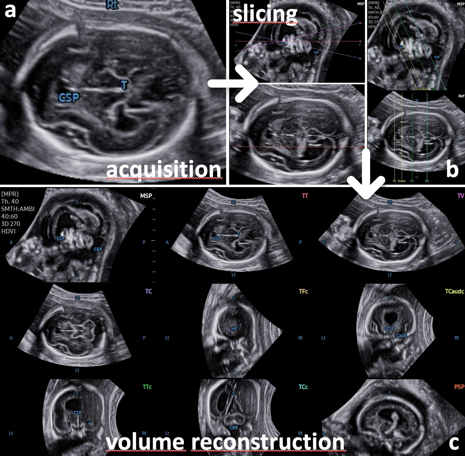
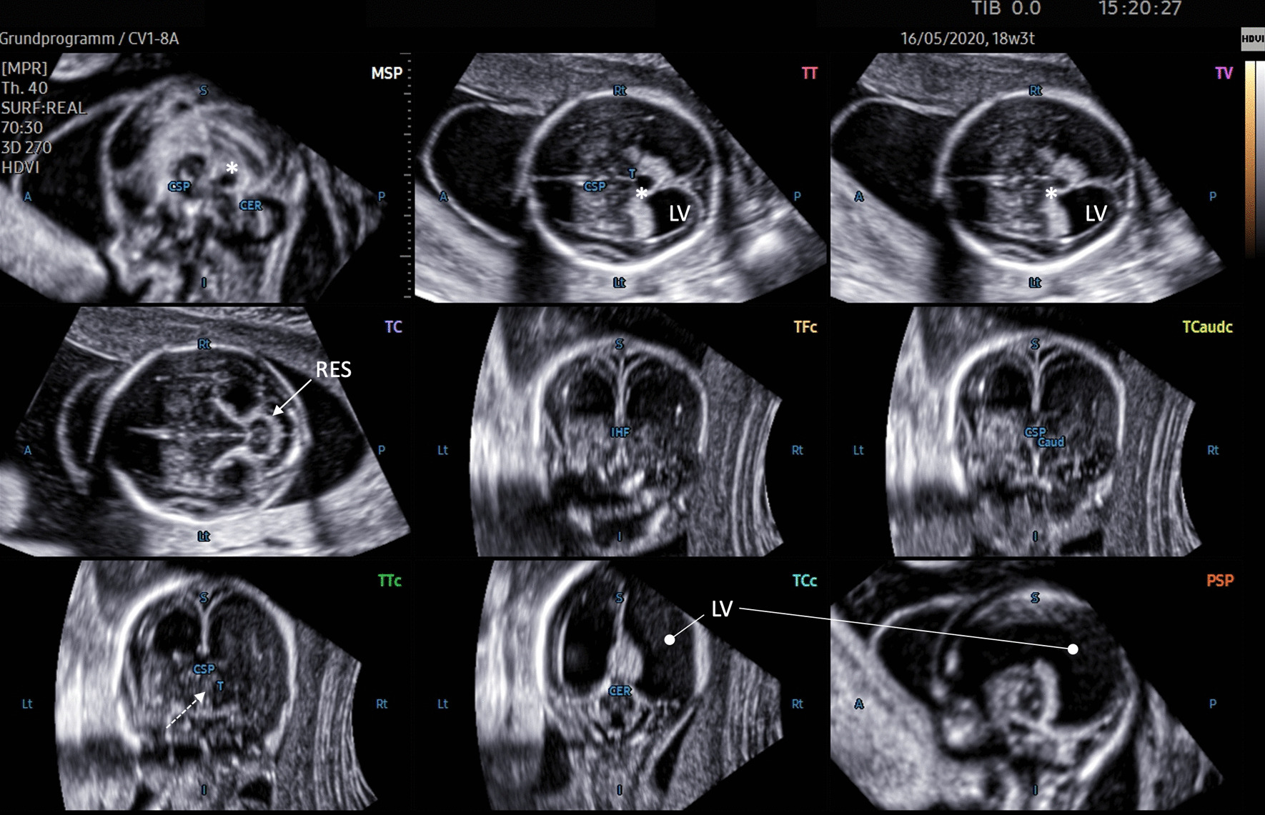
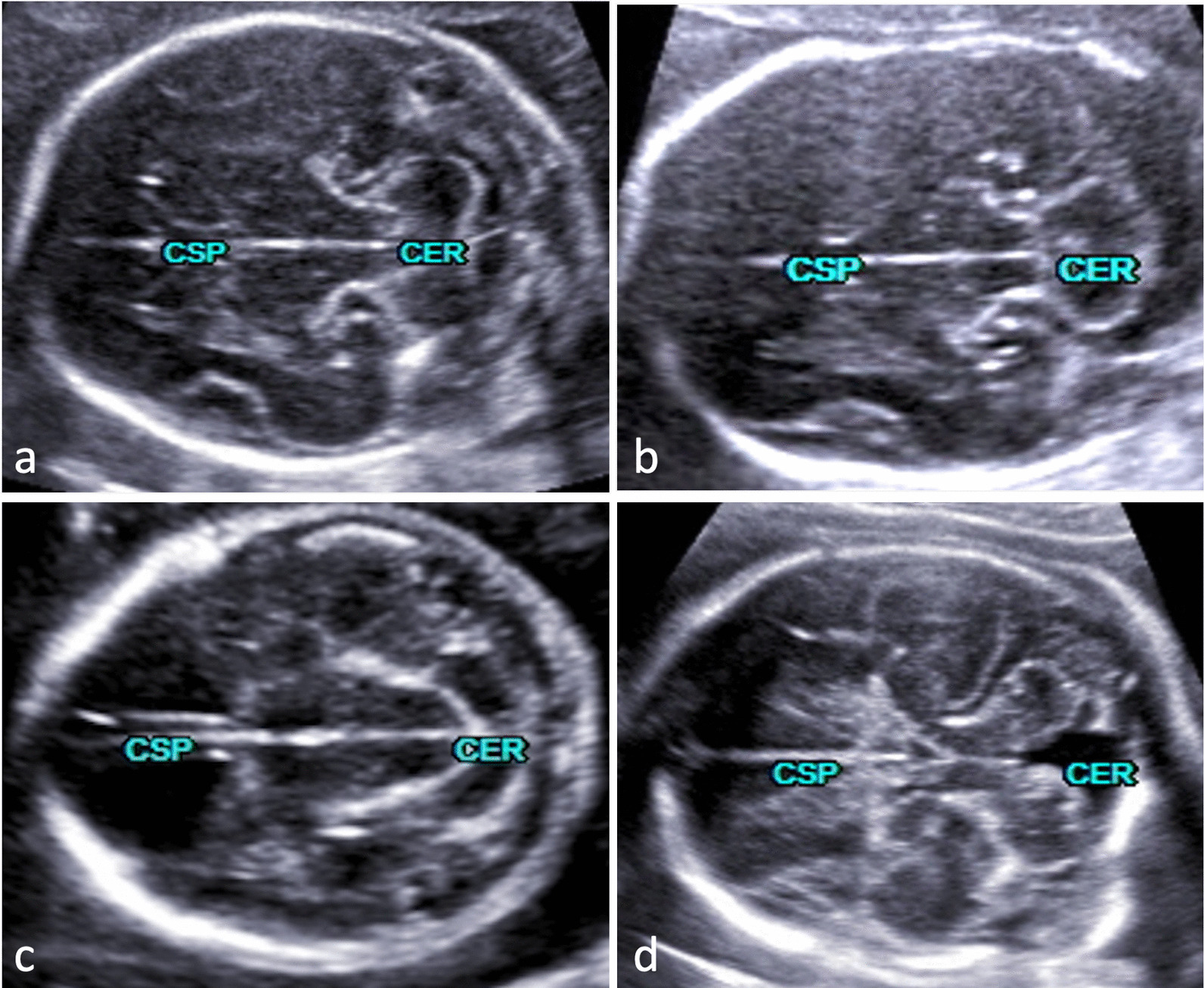
Methods: Stored 3D volumes extracted from a cohort of > 1.400 consecutive 2nd and 3rd trimester pregnancies (range 15-36 gestational weeks) were analyzed using the semiautomatic software tool 5D CNS+™, enabling detailed reconstruction of nine diagnostic planes of the fetal brain. All 3D data sets were examined and judged for plane accuracy, the need for manual adjustment, and fetal CNS anomalies affecting successful plane reconstruction.

Results: Based on our data of 91 fetuses with structural cerebral anomalies, we were able to reveal details of a wide range of CNS anomalies with application of the 5D CNS+™ technique. The corresponding anatomical features and consecutive changes of neighboring structures could be clearly demonstrated. Thus, a profound assessment of the entire altered CNS anatomy could be achieved in nearly all cases. The comparison with matched controls showed a significant difference in volume acquisition (p < 0.001) and in need for manual adjustment (p < 0.001) but not in the drop-out rates (p = 0.677) of both groups.

Conclusion: 5D CNS+™ is applicable in the majority of cases with brain lesions and constitutes a reliable tool even if the integrity of the fetal CNS is compromised by structural anomalies. Using volume data that were acquired in identical cutting sections needed for conventional biometry allows for detailed anatomic surveys grossly independent of the examiner's experience.

Keywords: 3D ultrasound; Anomalies; Brain; Central nervous system; Semiautomatic reconstruction.


Conflict of interest statement

The authors J. Weichert and M. Gembicki have received lecture fees from Samsung HME. A. Welp, Ch. Dracopoulos, J. L. Scharf and A. Rody declare that they have no conflict of interests.

Figures

Fig. 1
Fig. 1
The transthalamic diagnostic plane (recommended for biparietal diameter quantification) is needed for proper volume acquisition. The part of the frontal lobe is not sufficiently delineated, and an enlargement of the lateral ventricle is suspected. For further assessment, additional views are necessary. Application of the 5D CNS+™ algorithm automatically reslices the volume data set in predefined diagnostic planes as needed for a complete neurosonogram. The reconstructed planes show abnormal CNS morphology found in errors of ventral induction (e.g., semilobar holoprosencephaly)
Fig. 2
Fig. 2
Comparative alignment of complete neurosonograms comprising nine diagnostic planes of normal (a) and abnormal CNS anatomy (be). Panel b demonstrates a cystic lesion located in the midline. The information gained from different cutting sections shows a slight enlargement of the 3rd ventricle (and reduced interthalamic adhesion diameter) but normal appearance of the aqueduct seen in the midsagittal and transventricular and transcerebellar planes. The lesion is more caudally located, expanding symmetrically toward the median border of the lateral ventricles, both of which are slightly enlarged, suggesting a functional obstruction of cerebrospinal fluid (CSF) drainage via the left and right foramen of Monro. Panels c-e depict varying degrees of ventricular enlargement caused by different underlying causes. Agenesis of corpus callosum with colpocephaly (panel c), note the absent cavum septi pellucidi seen in transthalamic and anterior coronal planes; the patent aqueduct in the midsagittal and axial planes as well as the steer horn/bull’s head appearance of the anterior horns displayed in the transcaudate cutting section. Panel d shows features of occlusive hydrocephaly clearly emphasized in nearly all diagnostic planes and most likely caused by aqueductal stenosis (dilated 3rd ventricle and nonvisualization of the sonolucent aqueduct in midsagittal and axial planes). Panel e illustrates abnormal intracerebral findings attributed to a Chiari II malformation as a sequela from spina bifida aperta (descent of the tonsils and abnormal bowing of cerebellum in midsagittal and transcerebellar planes). There was also a marked dilatation of the lateral ventricles seen in all planes
Fig. 3
Fig. 3
Nine-image template after 5D CNS+™ application depicting abnormal CNS anatomy of a dichorial twin gestation at 18 completed weeks. The transthalamic plane (TT; acquisition plane) shows enlarged lateral ventricles (LV) and a fluid-filled area (*) in the midline (also seen in the sagittal and transventricular (TV) cutting sections), most likely representing a dilated suprapineal recessus. Turricephaly was clearly displayed in the sagittal planes. The aqueduct of Sylvius cannot be distinguished in either the sagittal or transcerebellar plane (TC), which accomplishes the clinical picture of an obstructed liquor circulation. The transverse diameter of the cerebellum is small, suggesting severe hypoplasia and fusion of the hemispheres, as found in rhombencephalosynapsis (RES; solid arrow). The coronal transthalamic plane (TTc) reveals thalamic fusion (dotted arrow)
Fig. 4
Fig. 4
Transcerebellar plane depicting different cerebellar appearances from normal (a) to abnormal (bd). The latter findings are part of gross intracranial pathology that needs further planes for delineating additional anomalies and establishing the final diagnosis. The rhombencephalosynapsis seen in panel b should necessarily stress an assessment of the ventricular system including the aqueduct of Sylvius (see also Fig. 3). In this particular case, aqueductal stenosis and triventricular enlargement were confirmed. An obstructed CSF pathway resulting in dilated lateral ventricles seen in all diagnostic planes underscored the impression of a Chiari II malformation (panel c). The fetus in panel d had vermian hypoplasia referred to as Dandy-Walker malformation
Fig. 5
Fig. 5
Intraventricular hemorrhage delineated using 5D CNS+™ in utero at 35 gestational weeks and correlated to the corresponding MR appearance on the 1st day after cesarean section. Note the severe hydrocephalus and the echogenic outlining of the ventricles. The blood clots predominantly seen in the axial and anterior coronal planes during prenatal imaging were markedly degraded in size or could not be reproduced postnatally. However, severe asymmetric enlargement of the ventricular system remained, and the cerebral cortex was thinned, corroborating the prenatal findings. MR images that correspond exactly for both the transcerebellar and parasagittal planes from 5D CNS+™ reconstruction could not be assigned due to the predetermined oblique cutting section.

References

    1. Garne E, Loane M, Addor MC, Boyd PA, Barisic I, Dolk H. Congenital hydrocephalus–prevalence, prenatal diagnosis and outcome of pregnancy in four European regions. Eur J Paediatr Neurol. 2010;14(2):150–155. doi: 10.1016/j.ejpn.2009.03.005. - DOI - PubMed
    1. Kinsner-Ovaskainen A, Morris JK, Garne E, Loane M, Lanzoni M. European monitoring of congenital anomalies: JRC-EUROCAT report on statistical monitoring of congenital anomalies (2008–2017) In Luxembourg. 2020 doi: 10.2760/65886. - DOI
    1. EUROCAT registry. Prevalence charts and tables. European Commission. European Platform on Rare Disease Registration Web site. https://eu-rd-platform.jrc.ec.europa.eu/eurocat/eurocat-data/prevalence_en. Updated 28.05.2021. Accessed 30.06.2021, 2021.
    1. Morris JK, Wellesley DG, Barisic I, et al. Epidemiology of congenital cerebral anomalies in Europe: a multicentre, population-based EUROCAT study. Arch Dis Child. 2019;104(12):1181–1187. doi: 10.1136/archdischild-2018-316733. - DOI - PubMed
    1. Van den Veyver IB. Prenatally diagnosed developmental abnormalities of the central nervous system and genetic syndromes: a practical review. Prenat Diagn. 2019;39(9):666–678. doi: 10.1002/pd.5520. - DOI - PubMed

Publication types

LinkOut - more resources