. 2022 Sep 2;17(1):57.
doi: 10.1186/s13024-022-00560-w.

LRP1 is a neuronal receptor for α-synuclein uptake and spread

Affiliations

LRP1 is a neuronal receptor for α-synuclein uptake and spread

Kai Chen et al. Mol Neurodegener. .

Abstract

Background: The aggregation and spread of α-synuclein (α-Syn) protein and related neuronal toxicity are the key pathological features of Parkinson's disease (PD) and Lewy body dementia (LBD). Studies have shown that pathological species of α-Syn and tau can spread in a prion-like manner between neurons, although these two proteins have distinct pathological roles and contribute to different neurodegenerative diseases. It is reported that the low-density lipoprotein receptor-related protein 1 (LRP1) regulates the spread of tau proteins; however, the molecular regulatory mechanisms of α-Syn uptake and spread, and whether it is also regulated by LRP1, remain poorly understood.

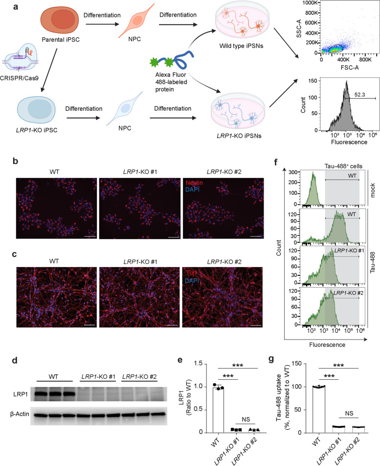
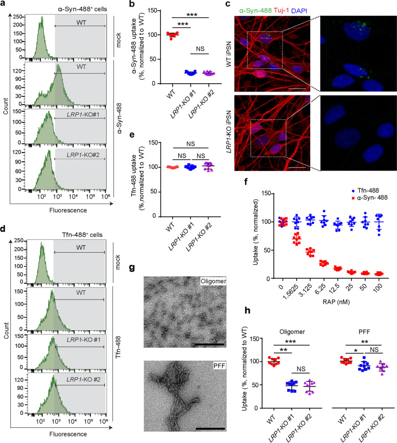
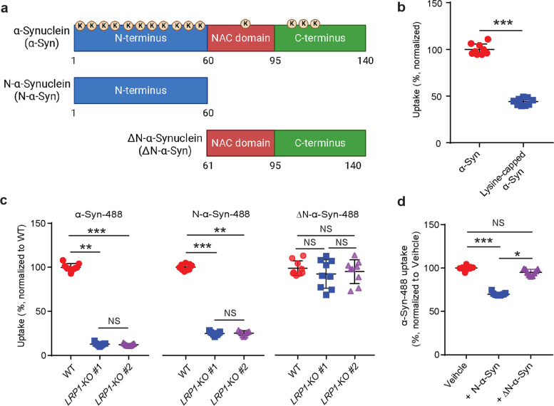
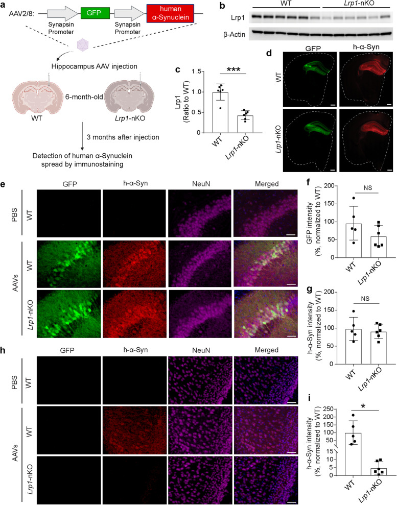
Methods: We established LRP1 knockout (LRP1-KO) human induced pluripotent stem cells (iPSCs) isogenic lines using a CRISPR/Cas9 strategy and generated iPSC-derived neurons (iPSNs) to test the role of LRP1 in α-Syn uptake. We treated the iPSNs with fluorescently labeled α-Syn protein and measured the internalization of α-Syn using flow cytometry. Three forms of α-Syn species were tested: monomers, oligomers, and pre-formed fibrils (PFFs). To examine whether the lysine residues of α-Syn are involved in LRP1-mediated uptake, we capped the amines of lysines on α-Syn with sulfo-NHS acetate and then measured the internalization. We also tested whether the N-terminus of α-Syn is critical for LRP1-mediated internalization. Lastly, we investigated the role of Lrp1 in regulating α-Syn spread with a neuronal Lrp1 conditional knockout (Lrp1-nKO) mouse model. We generated adeno-associated viruses (AAVs) that allowed for distinguishing the α-Syn expression versus spread and injected them into the hippocampus of six-month-old Lrp1-nKO mice and the littermate wild type (WT) controls. The spread of α-Syn was evaluated three months after the injection.

Results: We found that the uptake of both monomeric and oligomeric α-Syn was significantly reduced in iPSNs with LRP1-KO compared with the WT controls. The uptake of α-Syn PFFs was also inhibited in LRP1-KO iPSNs, albeit to a much lesser extent compared to α-Syn monomers and oligomers. The blocking of lysine residues on α-Syn effectively decreased the uptake of α-Syn in iPSNs and the N-terminus of α-Syn was critical for LRP1-mediated α-Syn uptake. Finally, in the Lrp1-nKO mice, the spread of α-Syn was significantly reduced compared with the WT littermates.

Conclusions: We identified LRP1 as a key regulator of α-Syn neuronal uptake, as well as an important mediator of α-Syn spread in the brain. This study provides new knowledge on the physiological and pathological role of LRP1 in α-Syn trafficking and pathology, offering insight for the treatment of synucleinopathies.

Keywords: Human induced pluripotent stem cells; Lewy body dementia; Low-density lipoprotein receptor-related protein 1; Parkinson’s disease; α-Synuclein.


Conflict of interest statement

G.B. consults for SciNeuro, has consulted for AbbVie, E-Scape, Eisai, and Vida Ventures and is on the scientific advisory board for Kisbee Therapeutics. D.M.H. is as an inventor on a patent licensed by Washington University to C2N Diagnostics on the therapeutic use of anti-tau antibodies. D.M.H. co-founded and is on the scientific advisory board of C2N Diagnostics. D.M.H. is on the scientific advisory board of Genentech, Denali and Cajal Neuroscience and consults for Alector. All other authors declare no competing interests.

Figures

Fig. 1
Fig. 1
Generation and validation of human induced pluripotent stem cells (iPSCs)-derived neurons (iPSNs) with LRP1 gene knockout (LRP1-KO). a Schematic diagram of the workflow for LRP1-KO iPSC generation, neural differentiation, and protein uptake assays. LRP1-KO iPSC colonies were obtained using CRISPR/Cas9 gene editing stratagy. Neural progenitor cells (NPCs) were then induced from the iPSCs and further differentiated into iPSNs. On day 14 to 16 of iPSN differentiation, neurons were treated with fluorescently labeled proteins and the uptake was measured by flow cytometry. b Immunofluorescence images showing NPCs from all three lines (WT, LRP1-KO#1, and LRP1-KO#2) were positive for neural precursor marker, Nestin. Scale bars, 100 μm. c, Immunofluorescence images of iPSNs from all three cell lines were positive for neuronal marker, Tuj1. Scale bars, 100 μm. d and e, Detection and quantification of LRP1 protein levels in WT and LRP1-KO iPSNs via Western blotting. f and g, Endocytosis of human tau in WT and LRP1-KO iPSNs measured by flow cytometry (100 nM, 3 h of treatment). Experiments in (f and g) were performed in technical duplicates or triplicates over three independent experiments. All data are expressed as mean ± s.d. with individual data points shown. Data were analyzed by One-way ANOVA with Tukey’s multiple comparisons test. NS, not significant; ***P < 0.001
Fig. 2
Fig. 2
LRP1 regulates α-Syn uptake in iPSNs. a and b, α-Syn uptake in WT and LRP1-KO iPSNs measured by flow cytometry (100 nM, 3 h of treatment). c Representative images of WT or LRP1-KO iPSNs after α-Syn uptake. Scale bars, 20 μm. d and e, Transferrin (Tfn) uptake in WT and LRP1-KO iPSNs measured by flow cytometry (300 nM, 3 h of treatment). f Uptake of α-Syn and Tfn in the presence of increasing concentrations of RAP. g EM images showing the structure of α-Syn oligomers and preformed fibrils (PFFs) used in panel h. Scale bars, 200 nm. h Uptake of α-Syn oligomers and PFFs in WT and LRP1-KO iPSNs (100 nM monomer equivalent, 3 h of treatment). All experiments in (a, b, d, e, f and h) were performed in technical duplicates or triplicates over three independent experiments. All data are expressed as mean ± s.d. with individual data points shown. Data were analyzed by One-way ANOVA with Tukey’s multiple comparisons test. NS, not significant; *P < 0.05, **P < 0.01, ***P < 0.001
Fig. 3
Fig. 3
LRP1 regulates α-Syn uptake via lysine residues in the N-terminus of α-Syn. a Schematic diagram of α-Syn domains highlighting the lysine residues (K). b Uptake of α-Syn and lysine-capped α-Syn in WT iPSNs. c, Uptake of α-Syn-488, N-α-Syn-488 and ΔN-α-Syn-488 in WT and LRP1-KO iPSNs. d Uptake of α-Syn-488 in the presence of excessive non-labeled α-Syn N-terminus (N-α-Syn) or α-Syn lacking N-terminus (ΔN-α-Syn) fragments. All experiments in (bd) were performed in technical duplicates or triplicates over three independent experiments. All data are expressed as mean ± s.d. with individual data points shown. Data in (b) was analyzed by unpaired two-sided t-test. ***P < 0.001. Data in (c and d) were analyzed by One-way ANOVA with Tukey’s multiple comparisons test. NS, not significant; *P < 0.05, **P < 0.01, ***P < 0.001
Fig. 4
Fig. 4
Neuronal Lrp1 knockout reduces α-Syn spread in vivo. a Schematic drawing for the stereotactic injection of AAV-synapsin-GFP-synapsin-h-α-Synuclein into the neuronal Lrp1 knockout (Lrp1-nKO) mice and wild type (WT) littermate controls, and the experimental workflow. b and c, Western blotting showing the endogenous Lrp1 protein in the cortex of WT (n = 6) and Lrp1-nKO mice (n = 6). d Representative sections showing GFP and h-α-Syn signals in mouse brains. Dotted line marks the outline of each section. Scale bars, 500 μm. e Representative images showing GFP and h-α-Syn signals in the hippocampus region from WT and Lrp1-nKO mice. Scale bars, 50 μm. f Quantitative analysis of GFP intensity in hippocampus from WT or Lrp1-nKO mice. g Quantitative analysis of h-α-Syn immunofluorescence intensity in hippocampus from WT or Lrp1-nKO mice. h Representative images showing h-α-Syn spreading to the cortex region from WT or Lrp1-nKO mice. Scale bars, 50 μm. i Quantitative analysis of h-α-Syn immunofluorescence intensity in the cortex from WT or Lrp1-nKO mice. Experiments in (ei) n = 5 mice (3 males and 2 females) for WT and n = 6 mice (3 males and 3 females) for Lrp1-nKO mice. All data are expressed as mean ± s.d. with individual data points shown. Data were analyzed by unpaired two-sided t-test. NS, not significant; *P < 0.05, ***P < 0.001

References

    1. Lashuel HA, Overk CR, Oueslati A, Masliah E. The many faces of alpha-synuclein: from structure and toxicity to therapeutic target. Nat Rev Neurosci. 2013;14:38–48. doi: 10.1038/nrn3406. - DOI - PMC - PubMed
    1. Meade RM, Fairlie DP, Mason JM. Alpha-synuclein structure and Parkinson’s disease - lessons and emerging principles. Mol Neurodegener. 2019;14:29. - PMC - PubMed
    1. Burre J, Sharma M, Tsetsenis T, Buchman V, Etherton MR, Sudhof TC. Alpha-synuclein promotes SNARE-complex assembly in vivo and in vitro. Science (New York, NY) 2010;329:1663–1667. doi: 10.1126/science.1195227. - DOI - PMC - PubMed
    1. Nemani VM, Lu W, Berge V, Nakamura K, Onoa B, Lee MK, Chaudhry FA, Nicoll RA, Edwards RH. Increased expression of α-Synuclein reduces neurotransmitter release by inhibiting synaptic vesicle reclustering after endocytosis. Neuron. 2010;65:66–79. doi: 10.1016/j.neuron.2009.12.023. - DOI - PMC - PubMed
    1. Scott D, Roy S. α-Synuclein inhibits intersynaptic vesicle mobility and maintains recycling-pool homeostasis. J Neurosci. 2012;32:10129–10135. doi: 10.1523/JNEUROSCI.0535-12.2012. - DOI - PMC - PubMed