. 2022 Sep 2;53(1):67.
doi: 10.1186/s13567-022-01084-3.

Evaluation of alpaca tracheal explants as an ex vivo model for the study of Middle East respiratory syndrome coronavirus (MERS-CoV) infection

Affiliations

Evaluation of alpaca tracheal explants as an ex vivo model for the study of Middle East respiratory syndrome coronavirus (MERS-CoV) infection

Nigeer Te et al. Vet Res. .

Abstract

Middle East respiratory syndrome coronavirus (MERS-CoV) poses a serious threat to public health. Here, we established an ex vivo alpaca tracheal explant (ATE) model using an air-liquid interface culture system to gain insights into MERS-CoV infection in the camelid lower respiratory tract. ATE can be infected by MERS-CoV, being 103 TCID50/mL the minimum viral dosage required to establish a productive infection. IFNs and antiviral ISGs were not induced in ATE cultures in response to MERS-CoV infection, strongly suggesting that ISGs expression observed in vivo is rather a consequence of the IFN induction occurring in the nasal mucosa of camelids.

Keywords: Air-liquid interface; MERS-CoV; alpaca; camelid; ex vivo model; tracheal explants.


Conflict of interest statement

The authors declare that they have no competing interests.

Figures

Figure 1
Figure 1
Schematic diagram of the experimental time course and settings of the air-liquid (ALI) device for the alpaca tracheal explant (ATE) model. (A) A schematic overview of the ALI interface set-up and (B) a photo of an ATE (arrow) resting onto an inox gauze within a 6-well culture plate. (C) General overview of the experiment indicating the time points in which explants were collected, cultured, infected, and sampled. Upon necropsy, ATE were isolated from alpacas (n = 3) and cultured in an ALI interface system. ATE were apically infected after 24 h of culture with MERS-CoV Qatar15/2015 strain at three different doses: 102, 103, and 104 TCID50/mL. Explant replicates from each animal were collected for virological, pathological, and immunological assessments at -24, 0, 24, 48 and 72 h respectively.
Figure 2
Figure 2
Evaluation of alpaca tracheal explants (ATE) thickness after ex vivo culture by means of light photomicrographs. The trachea was harvested upon necropsy, followed by infection at different doses of MERS-CoV (right of the figure). ATE were collected at different time points and were fixed in 10% neutral-buffered formalin. Sections of each formalin-fixed paraffin embedded ATE were stained by hematoxylin-eosin to evaluate the thickness of the epithelium by measuring five randomly selected fields across each section (indicated with a black arrow at the left) at –24, 0, 24, 48 and 72 hpi, respectively.
Figure 3
Figure 3
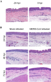
Histopathological study of alpaca tracheal explants (ATE). Hematoxylin-eosin staining was applied to assess histological alterations of ATE (A) prior and (B) after MERS-CoV infection. No significant lesions were observed in any of the studied explants, inoculated or not with MERS-CoV. Original magnification: × 200 for all tissues.
Figure 4
Figure 4
MERS-CoV loads in alpaca tracheal explants (ATE). (A) MERS-CoV genomic (left) and subgenomic (right) RNA was determined by microfluidic RT-qPCR assays (Sheet A in Additional file 1). Each line represents the mean Cq value and error bars display standard deviation intervals of ATE from 3 animals sampled at 0, 24, 48 and 72 hpi, respectively. Blue, purple, and red dots indicate ATE infected with 102, 103 and 104 TCID50/mL MERS-CoV, respectively. (B) Tissue tropism of MERS-CoV in ATE. At 72 hpi, MERS-CoV N protein (brown staining) was detected by immunohistochemistry in the epithelium of ATE that were infected with 103 and 104 TCID50/mL doses respectively; original magnification: × 200 for both tissues. ATE, alpaca tracheal explants; 1/3 and 2/3 indicate the detection of viral RNA in one out of three or two out of three explants, respectively.
Figure 5
Figure 5
Kinetics of innate immune gene profiles of ATE in response to different MERS-CoV infectious doses. Total RNA was extracted from ATE, followed by conversion to cDNA. The Fluidigm Biomark microfluidic assay was used to quantify transcripts from innate immune genes at different hpi. After normalization, fold change values between controls (freshly prepared explants) and mock or infected ATE were calculated. The resulting heatmap shows color variations corresponding to log2 fold change values, blue for increased and brown for decreased gene expression, respectively. The khaki rectangles indicate no expression of the corresponding gene. TFs, transcription factors; CKs, chemokines; ADs, adaptors; RT, receptor.

References

    1. Zaki AM, van Boheemen S, Bestebroer TM, Osterhaus ADME, Fouchier RAM. Isolation of a novel coronavirus from a man with pneumonia in Saudi Arabia. N Engl J Med. 2012;367:1814–1820. doi: 10.1056/nejmoa1211721. - DOI - PubMed
    1. Arabi YM, Arifi AA, Balkhy HH, Najm H, Aldawood AS, Ghabashi A, Hawa H, Alothman A, Khaldi A, Al Raiy B. Clinical course and outcomes of critically ill patients with Middle East respiratory syndrome coronavirus infection. Ann Intern Med. 2014;160:389–397. doi: 10.7326/M13-2486. - DOI - PubMed
    1. Assiri A, Al-Tawfiq JA, Al-Rabeeah AA, Al-Rabiah FA, Al-Hajjar S, Al-Barrak A, Flemban H, Al-Nassir WN, Balkhy HH, Al-Hakeem RF, Makhdoom HQ, Zumla AI, Memish ZA. Epidemiological, demographic, and clinical characteristics of 47 cases of Middle East respiratory syndrome coronavirus disease from Saudi Arabia: a descriptive study. Lancet Infect Dis. 2013;13:752–761. doi: 10.1016/S1473-3099(13)70204-4. - DOI - PMC - PubMed
    1. Zumla A, Hui DS, Perlman S. Middle East respiratory syndrome. Lancet. 2015;386:995–1007. doi: 10.1016/S0140-6736(15)60454-8. - DOI - PMC - PubMed
    1. WHO | Middle East respiratory syndrome coronavirus (MERS-CoV). https://www.who.int/emergencies/mers-cov/en/. Accessed 18 May 2022

Substances