Review
. 2023 Jan;95(1):e28122.
doi: 10.1002/jmv.28122. Epub 2022 Sep 13.

COVID-19 immunopathology: From acute diseases to chronic sequelae

Affiliations
Review

COVID-19 immunopathology: From acute diseases to chronic sequelae

Mohd Arish et al. J Med Virol. 2023 Jan.

Abstract

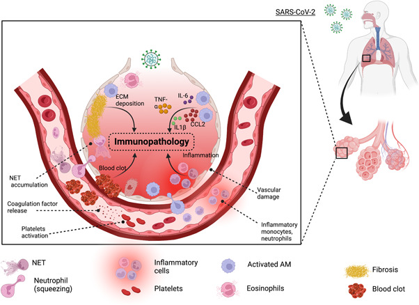
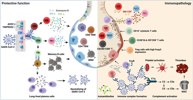
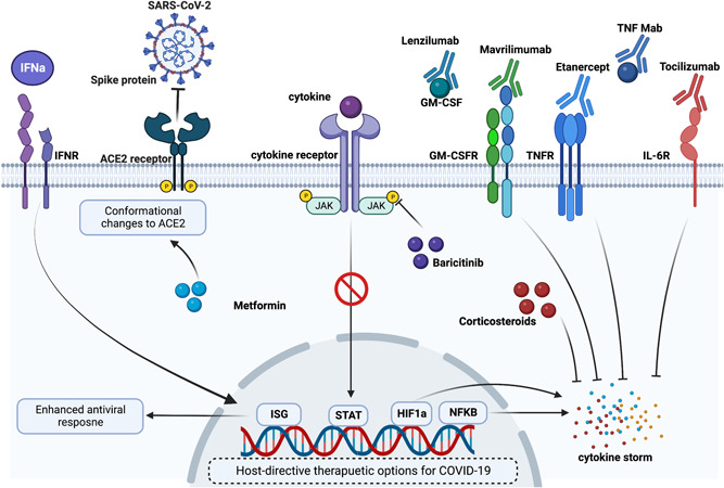
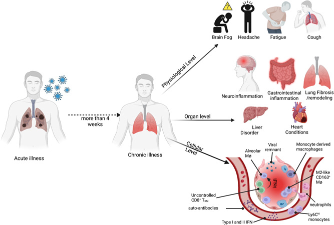
The clinical manifestation of coronavirus disease 2019 (COVID-19) mainly targets the lung as a primary affected organ, which is also a critical site of immune cell activation by severe acute respiratory syndrome coronavirus 2 (SARS-CoV-2). However, recent reports also suggest the involvement of extrapulmonary tissues in COVID-19 pathology. The interplay of both innate and adaptive immune responses is key to COVID-19 management. As a result, a robust innate immune response provides the first line of defense, concomitantly, adaptive immunity neutralizes the infection and builds memory for long-term protection. However, dysregulated immunity, both innate and adaptive, can skew towards immunopathology both in acute and chronic cases. Here we have summarized some of the recent findings that provide critical insight into the immunopathology caused by SARS-CoV-2, in acute and post-acute cases. Finally, we further discuss some of the immunomodulatory drugs in preclinical and clinical trials for dampening the immunopathology caused by COVID-19.

Keywords: SARS coronavirus; immnopathology; immune responses; respiratory tract.


Conflict of interest statement

Jie Sun is a consultant for TeneoFour company.

Figures

FIGURE 1
FIGURE 1
Innate cell‐mediated immunopathology in COVID‐19: Upon viral entry, there is a cascade of events that leads to inflammation, vascular damage, and blot. Tissue‐resident alveolar macrophages (AMs) and interstitial macrophages are among the first responders to SARS‐CoV‐2, which secret inflammatory cytokines including TNF, IL‐6, IL‐1β, and CCL2 that in addition to building up local inflammation but also attract monocyte and neutrophils to the site of infection. Furthermore, IL‐1β favors the expansion of pathological fibroblasts that further contribute to fibrosis. SARS‐CoV‐2 can also stimulate platelets and neutrophils to secrete coagulation factors resulting in the formation of leukocyte–platelet aggregates and NETs, respectively. Lastly, fibroblast proliferation leads to the deposition of extracellular matrix (ECM) and fibrin in alveolar space further complicating the lung alveolar structure. COVID‐19, coronavirus disease 2019; IL‐6, interleukin 6; NET, neutrophil extracellular trap; SARS‐CoV‐2, severe acute respiratory syndrome coronavirus 2; TNF, tumor necrosis factor.
FIGURE 2
FIGURE 2
The protective versus pathogenic adaptive immune responses in COVID‐19. Left: When SARS‐CoV‐2 invades the host respiratory tract, the viral antigen can be detected and presented by DCs to either CD4+ T or CD8+ T cells for their activation. Naïve CD4+ T cells mainly differentiate into T helper 1 (Th1) and T follicular helper cells (Tfh). Th1 cells possess antiviral effects by producing higher levels of IFN‐γ, TNF, and IL‐2. Tfh cells provide help to B cells for somatic hypermutations and affinity maturation of germinal center reactions to generate memory B cells and long‐lived antibody‐producing plasma cells. The viral‐specific antibodies secreted by plasma cells play a protective role by neutralizing the virus. Activated CD8+ T cells produce effector cytokines and cytotoxic molecules, including IFN‐γ, TNF, IL‐2, and granzyme B, controlling viral infections. After viral clearance, memory CD4+ T, CD8+ T, and B cells are developed in the circulation and lungs to protect against secondary infections. Right: Excessive T cell responses are associated with severe COVID‐19, including IL‐6‐ and GM‐CSF‐producing Th1 or Th17 cells, CD16+ cytotoxic T cells, CXCR6+ CD8+ T cells, as well as dysregulated Treg cells. On the other hand, the production of autoantibodies, the formation of immune complexes, and complement activation also contribute to the disease progression of COVID‐19. ACE2, angiotensin‐converting enzyme 2; BRM, resident memory B; COVID‐19, coronavirus disease 2019; DC, dendritic cell; FcγR, Fc‐γ receptor; GM‐CSF, granulocyte‐macrophage colony‐stimulating factor; IFN‐γ, interferon‐γ; IL‐2, interleukin 2; MAC, Membrane attack complex; SARS‐CoV‐2, severe acute respiratory syndrome coronavirus 2; TMPRSS2, transmembrane serine protease 2; TNF, tumor necrosis factor; Treg, regulatory T cell; TRM, tissue‐resident memory.
FIGURE 3
FIGURE 3
Potential immunomodulatory drugs for acute COVID‐19: Acute COVID‐19 is accompanied by hyperinflammatory responses and hence use of immunomodulatory in several clinical and preclinical settings has shown therapeutic benefits. Immunomodulatory drugs, such as metformin, corticosteroids, and baricitinib have shown reduced inflammation following SARS‐CoV‐2 infection. Additionally, monoclonal antibodies (mAbs) such as lenzilumab, mavrilimumab, etanercept, tocilizumab, and TNF mAb have been studied in various clinical trials for their beneficial role in dampening COVID‐19‐induced inflammation. As early ISG expression is required for effective viral clearance, treatment with recombinant IFNs has also been proposed to mitigate viral load. ACE2, angiotensin‐converting enzyme 2; COVID‐19, coronavirus disease 2019; GM‐CSF, granulocyte‐macrophage colony‐stimulating factor; GM‐CSFR, GM‐CSF receptor; HIF‐1a, hypoxia‐inducible factor 1a; IFN, interferon; IFNR, IFN receptor; IL‐6R, interleukin 6R; ISG, interferon‐stimulated gene;NF‐κB, nuclear factor‐κB; SARS‐CoV‐2, severe acute respiratory syndrome coronavirus 2; STAT, signal transducer and activator of transcription; TNF, tumor necrosis factor; TNFR, tumor necrosis factor receptor.
FIGURE 4
FIGURE 4
Immunopathology in long COVID: Immunopathology in long COVID is studied at different biological levels. At the physiological levels, individuals recovering from acute SARS‐CoV‐2 infection have been complaining about brain fog, headache, fatigue, cough, and so forth, for a prolonged period. At the organ levels, in infected patients, there have been reports of long‐term neuro‐and gastroinflammation. In some individuals, there have been incidences of liver and heart conditions. However, the cellular insight into this chronic illness remains poorly understood. Some of the recent reports have suggested the presence of viral remnants, prolonged systemic or tissue inflammatory responses, and/or the presence of autoantibodies may contribute to the disease etiology. COVID‐19, coronavirus disease 2019; IFN, interferon; SARS‐CoV‐2, severe acute respiratory syndrome coronavirus 2.

References

    1. COVID‐19 dashboard by the Center for Systems Science and Engineering (CSSE) at Johns Hopkins University (JHU). Accessed September 7, 2022. https://coronavirus.jhu.edu/map.html
    1. Harvey WT, Carabelli AM, Jackson B, et al. SARS‐CoV‐2 variants, spike mutations and immune escape. Nat Rev Microbiol. 2021;19(7):409‐424. - PMC - PubMed
    1. Lopes‐Pacheco M, Silva PL, Cruz FF, et al. Pathogenesis of multiple organ injury in COVID‐19 and potential therapeutic strategies. Front Physiol. 2021;12:593223. - PMC - PubMed
    1. Yang L, Liu S, Liu J, et al. COVID‐19: immunopathogenesis and immunotherapeutics. Signal Transduct Target Ther. 2020;5(1):128. - PMC - PubMed
    1. Huang W, Berube J, McNamara M, et al. Lymphocyte subset counts in COVID‐19 patients: a meta‐analysis. Cytometry A. 2020;97(8):772‐776. - PMC - PubMed

Publication types