Cathepsin F is a potential marker for senescent human skin fibroblasts and keratinocytes associated with skin aging
- PMID: 36057013
- PMCID: PMC9886782
- DOI: 10.1007/s11357-022-00648-7
Cathepsin F is a potential marker for senescent human skin fibroblasts and keratinocytes associated with skin aging
Abstract
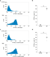
Cellular senescence is characterized by cell cycle arrest and the senescence-associated secretory phenotype (SASP) and can be triggered by a variety of stimuli, including deoxyribonucleic acid (DNA) damage, oxidative stress, and telomere exhaustion. Cellular senescence is associated with skin aging, and identification of specific markers of senescent cells is essential for development of targeted therapies. Cathepsin F (CTSF) has been implicated in dermatitis and various cancers and participates in cell immortalization through its association with Bcl family proteins. It is a candidate therapeutic target to specifically label and eliminate human skin fibroblasts and keratinocytes immortalized by aging and achieve skin rejuvenation. In this study, we investigated whether CTSF is associated with senescence in human fibroblasts and keratinocytes. In senescence models, created using replicative aging, ionizing radiation exposure, and the anticancer drug doxorubicin, various senescence markers were observed, such as senescence-associated β-galactosidase (SA-β-gal) activity, increased SASP gene expression, and decreased uptake of the proliferation marker BrdU. Furthermore, CTSF expression was elevated at the gene and protein levels. In addition, CTSF-positive cells were abundant in aged human epidermis and in some parts of the dermis. In the population of senescent cells with arrested division, the number of CTSF-positive cells was significantly higher than that in the proliferating cell population. These results suggest that CTSF is a candidate for therapeutic modalities targeting aging fibroblasts and keratinocytes.
Keywords: Cathepsin; Cellular senescence; Fibroblast; Keratinocytes; Senescence-associated secretory phenotype; Skin aging.
© 2022. The Author(s).
Conflict of interest statement
The authors declare no competing interests.
Figures


Similar articles
-
Identification of Apolipoprotein D as a Dermal Fibroblast Marker of Human Aging for Development of Skin Rejuvenation Therapy.Rejuvenation Res. 2023 Apr;26(2):42-50. doi: 10.1089/rej.2022.0056. Epub 2023 Mar 1. Rejuvenation Res. 2023. PMID: 36571249
-
Identification of a new human senescent skin cell marker ribonucleoside-diphosphate reductase subunit M2 B.Biogerontology. 2024 Nov;25(6):1239-1251. doi: 10.1007/s10522-024-10135-5. Epub 2024 Sep 11. Biogerontology. 2024. PMID: 39261410
-
Senotherapeutic-like effect of Silybum marianum flower extract revealed on human skin cells.PLoS One. 2021 Dec 16;16(12):e0260545. doi: 10.1371/journal.pone.0260545. eCollection 2021. PLoS One. 2021. PMID: 34914725 Free PMC article.
-
Aging of the cells: Insight into cellular senescence and detection Methods.Eur J Cell Biol. 2020 Aug;99(6):151108. doi: 10.1016/j.ejcb.2020.151108. Epub 2020 Jul 12. Eur J Cell Biol. 2020. PMID: 32800277 Review.
-
Flavonoids in Skin Senescence Prevention and Treatment.Int J Mol Sci. 2021 Jun 25;22(13):6814. doi: 10.3390/ijms22136814. Int J Mol Sci. 2021. PMID: 34201952 Free PMC article. Review.
Cited by
-
The Potential of Senescence as a Target for Developing Anticancer Therapy.Int J Mol Sci. 2023 Feb 8;24(4):3436. doi: 10.3390/ijms24043436. Int J Mol Sci. 2023. PMID: 36834846 Free PMC article. Review.
-
Exploring the role of cathepsins in sarcopenia-related traits: Insights from a Mendelian randomization study.Medicine (Baltimore). 2025 Jun 6;104(23):e42700. doi: 10.1097/MD.0000000000042700. Medicine (Baltimore). 2025. PMID: 40489878 Free PMC article.
-
Genetic alterations in coronary cell lines exposed to sirolimus and paclitaxel.Arch Toxicol. 2025 Jun 6. doi: 10.1007/s00204-025-04101-4. Online ahead of print. Arch Toxicol. 2025. PMID: 40481235
-
Cathepsins in oral diseases: mechanisms and therapeutic implications.Front Immunol. 2023 Jun 2;14:1203071. doi: 10.3389/fimmu.2023.1203071. eCollection 2023. Front Immunol. 2023. PMID: 37334378 Free PMC article. Review.
-
Cathepsins and their role in gynecological cancers: Evidence from two-sample Mendelian randomization analysis.Medicine (Baltimore). 2025 Mar 7;104(10):e41653. doi: 10.1097/MD.0000000000041653. Medicine (Baltimore). 2025. PMID: 40068078 Free PMC article.
References
Publication types
MeSH terms
Substances
LinkOut - more resources
Full Text Sources
Medical

