. 2022 Sep 3;10(1):128.
doi: 10.1186/s40478-022-01432-6.

LATE-NC aggravates GVD-mediated necroptosis in Alzheimer's disease

Affiliations

LATE-NC aggravates GVD-mediated necroptosis in Alzheimer's disease

Marta J Koper et al. Acta Neuropathol Commun. .

Abstract

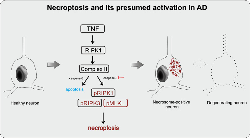
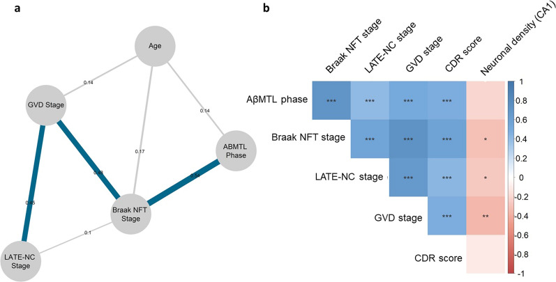
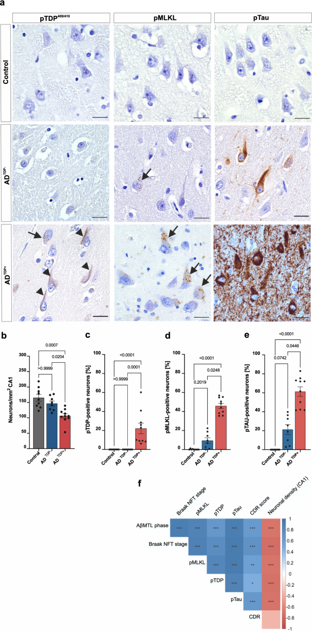
It has become evident that Alzheimer's Disease (AD) is not only linked to its hallmark lesions-amyloid plaques and neurofibrillary tangles (NFTs)-but also to other co-occurring pathologies. This may lead to synergistic effects of the respective cellular and molecular players, resulting in neuronal death. One of these co-pathologies is the accumulation of phosphorylated transactive-response DNA binding protein 43 (pTDP-43) as neuronal cytoplasmic inclusions, currently considered to represent limbic-predominant age-related TDP-43 encephalopathy neuropathological changes (LATE-NC), in up to 70% of symptomatic AD cases. Granulovacuolar degeneration (GVD) is another AD co-pathology, which also contains TDP-43 and other AD-related proteins. Recently, we found that all proteins required for necroptosis execution, a previously defined programmed form of neuronal cell death, are present in GVD, such as the phosphorylated necroptosis executioner mixed-lineage kinase domain-like protein (pMLKL). Accordingly, this protein is a reliable marker for GVD lesions, similar to other known GVD proteins. Importantly, it is not yet known whether the presence of LATE-NC in symptomatic AD cases is associated with necroptosis pathway activation, presumably contributing to neuron loss by cell death execution. In this study, we investigated the impact of LATE-NC on the severity of necroptosis-associated GVD lesions, phosphorylated tau (pTau) pathology and neuronal density. First, we used 230 human post-mortem cases, including 82 controls without AD neuropathological changes (non-ADNC), 81 non-demented cases with ADNC, i.e.: pathologically-defined preclinical AD (p-preAD) and 67 demented cases with ADNC. We found that Braak NFT stage and LATE-NC stage were good predictors for GVD expansion and neuronal loss in the hippocampal CA1 region. Further, we compared the impact of TDP-43 accumulation on hippocampal expression of pMLKL-positive GVD, pTau as well as on neuronal density in a subset of nine non-ADNC controls, ten symptomatic AD cases with (ADTDP+) and eight without LATE-NC (ADTDP-). Here, we observed increased levels of pMLKL-positive, GVD-exhibiting neurons in ADTDP+ cases, compared to ADTDP- and controls, which was accompanied by augmented pTau pathology. Neuronal loss in the CA1 region was increased in ADTDP+ compared to ADTDP- cases. These data suggest that co-morbid LATE-NC in AD impacts not only pTau pathology but also GVD-mediated necroptosis pathway activation, which results in an accelerated neuronal demise. This further highlights the cumulative and synergistic effects of comorbid pathologies leading to neuronal loss in AD. Accordingly, protection against necroptotic neuronal death appears to be a promising therapeutic option for AD and LATE.

Keywords: Cell death; Granulovacuolar degeneration; LATE-NC; Necroptosis; Protein aggregation; TDP-43; pMLKL; pTau.


Conflict of interest statement

MO served as consultant for Axon neuroscience and Fujirebio and gave invited talks for Roche and Fujirebio. CAFvA received honoraria from serving on the scientific advisory board of Nutricia GmbH (2014), Roche (2018) and Hong Kong University Research council (2014) and has received funding for travel and speaker honoraria from Nutricia GmbH (2014–2015), Lilly Deutschland GmbH (2013–2016), Desitin Arzneimittel GmbH (2014), Biogen (2016–2018), Roche (2017–2019) and Dr. Willmar Schwabe GmbH &Co. KG (2014–2019). BDS is or has been a consultant for Eli Lilly, Biogen, Janssen Pharmaceutica, Eisai, AbbVie and other companies. BDS is also a scientific founder of Augustine Therapeutics and a scientific founder and stockholder of Muna therapeutics. DRT received speaker honorary from Novartis Pharma AG (Switzerland) and Biogen (USA), travel reimbursement from GE-Healthcare (UK) and UCB (Belgium) and collaborated with Novartis Pharma AG (Switzerland), Probiodrug (Germany), GE-Healthcare (UK), and Janssen Pharmaceutical Companies (Belgium).

Figures

Fig. 1
Fig. 1
The necroptosis pathway and its presumed activation in AD. Necroptosis is a programmed form of cell death which starts with the activation of death receptors. RIPK1 is then recruited and self-phosphorylated [12]. The inhibition or activation of caspase-8 determines whether necroptosis or apoptosis takes place. If caspase-8 is inhibited, the necrosome complex is formed, which includes activated pRIPK1, pRIPK3 and pMLKL. This complex translocates to the cell membrane, resulting in membrane swelling and bursting mediated by pMLKL, the necroptosis executor [12]. This is followed by release of intracellular components, inflammatory response and ultimately neuronal death. Necroptosis activation has been previously linked to AD [12]. Here, the activated necrosome components pRIPK1, pRIPK3 and pMLKL accumulate in GVD granules presumably indicating an aberrant/delayed execution process of necroptosis in neurons with GVD [41]
Fig. 2
Fig. 2
Tau, LATE-NC and GVD expansion are strongly associated. a Network constructed with graphical LASSO reveals that GVD is strongly associated with Braak NFT stage and mediates its relationship with LATE-NC stage (n = 221). The presence of connecting lines, i.e., edges between nodes (variables) indicates a positive and non-spurious partial correlation coefficient (with r values displayed on the edges and represented by the width of an edge). Edges representing correlation coefficients higher than 0.4 are marked in blue. b Semi-partial correlation matrix adjusted for sex shows that LATE-NC and Braak NFT stages are significantly correlated with GVD stage and CDR score, and that Braak NFT, LATE-NC and GVD stages negatively correlates with CA1-hippocampus neuronal density (n = 64). The p-values were adjusted with the Holm–Bonferroni method
Fig. 3
Fig. 3
TDP-43 and pTau are co-expressed in necroptosis-positive neurons. Triple labeling immunofluorescence with antibodies against pTDP-43 (S409/S410), pTau (S202/T205) and pMLKL (S358) in the CA1 sub-hippocampal field of an ADTDP+ case. Nuclei are stained with Hoechst solution. Of note, a pTDP-43 and pMLKL are co-localized in some GVD granules (arrows) and b pTDP-43 and pTau co-localize in nearby neurons (arrowheads). We exclude the signal observed being considered as unspecific lipofuscin because such signal usually shows in the blue (Hoeschst) channel as orange granules, which in this case is absent
Fig. 4
Fig. 4
pMLKL and pTau severity as well as neuronal loss are increased in AD cases with LATE-NC. a Local accumulation of phosphorylated tau and MLKL is observed in ADTDP+. DAB immunohistochemical staining of pTDP-43 (S409/S410), pTau (S202/T205) and pMLKL (S358) in the CA1-subiculum field of a control, ADTDP− and ADTDP+ case (cases 7, 16 and 21 are displayed, see Table 2). Scale bars = 50 µm. b ADTDP+ cases display decreased neuronal density compared to controls and ADTDP− and controls. Quantitative data representing the number of total neurons per mm2 per group in the CA1 subfield of the hippocampus. Quantification of the number of CA1 positive neurons for c pTDP-43 d pMLKL and e pTau. ADTDP+ cases display significantly higher severity of pMLKL and pTau lesions. Data are presented as mean ± SEM. N = 27 (controls = 9, ADTDP− = 8, ADTDP+  = 10). f Partial Spearman correlation matrix controlled for age and multiple comparisons (Holm-Bonferroni test) in this cohort confirms that the accumulation of pTDP-43, pMLKL and pTau pathologies are significantly correlated with neuronal density in the CA1, AβMTL phase and Braak NFT stage. Supporting images showing overviews of the whole hippocampus of cases 12, 16, 21 and 22 (Table 2) stained with pMLKL can be found in the public repository BioImage Archive, with the hyperlink: https://www.ebi.ac.uk/biostudies/studies/S-BIAD514?key=475a3bbe-6fc9-476e-8e45-6429422b85bf
Fig. 5
Fig. 5
Proposed TDP-43, pTau and pMLKL interplay in AD. Aβ and TDP-43 pathologies accelerate pTau pathology in AD (black and orange arrows, respectively). We also hypothesize that TDP-43 exacerbates necrosome-positive GVD lesions through pTau (blue arrow), giving rise to neurons bearing tangles, pTDP-43 inclusions and pMLKL-positive GVD. This eventually leads to neurodegeneration and neuronal loss

References

    1. Alzheimer’s Association (2021) 2021 Alzheimer’s Disease facts and figures. Alzheimers Dement 17:327–406 - PubMed
    1. Arriagada PV, Growdon JH, Hedley-Whyte ET, Hyman BT. Neurofibrillary tangles but not senile plaques parallel duration and severity of Alzheimer’s disease. Neurology. 1992;42:631–639. doi: 10.1212/WNL.42.3.631. - DOI - PubMed
    1. Attems J, Jellinger K. Neuropathological correlates of cerebral multimorbidity. Curr Alzheimer Res. 2013;10:569–577. doi: 10.2174/15672050113109990002. - DOI - PubMed
    1. Attems J, Neltner JH, Nelson PT. Quantitative neuropathological assessment to investigate cerebral multi-morbidity. Alzheimers Res Ther. 2014;6:85. doi: 10.1186/s13195-014-0085-y. - DOI - PMC - PubMed
    1. Ball MJ. Neuronal loss, neurofibrillary tangles and granulovacuolar degeneration in the hippocampus with ageing and dementia. Acta Neuropathol. 1977;37:111–118. doi: 10.1007/BF00692056. - DOI - PubMed

Publication types

Substances